ARG42328
anti-CD1c antibody [L161] (APC)
anti-CD1c antibody [L161] (APC) for Flow cytometry and Human
Overview
| Product Description | APC-conjugated Mouse Monoclonal antibody [L161] recognizes CD1c |
|---|---|
| Tested Reactivity | Hu |
| Tested Application | FACS |
| Specificity | The mouse monoclonal antibody L161 recognizes an extracellular epitope of CD1c, (R7), a 43 kDa type I glycoprotein associated with beta2-microglobulin. It is expressed on cortical thymocytes (strongly), Langerhans cells, dendritic cells, B and some T cells. |
| Host | Mouse |
| Clonality | Monoclonal |
| Clone | L161 |
| Isotype | IgG1, kappa |
| Target Name | CD1c |
| Antigen Species | Human |
| Immunogen | Human thymocytes. |
| Conjugation | APC |
| Alternate Names | R7; CD antigen CD1c; CD1A; CD1; T-cell surface glycoprotein CD1c; BDCA1 |
Application Instructions
| Application Suggestion |
|
||||
|---|---|---|---|---|---|
| Application Note | * The dilutions indicate recommended starting dilutions and the optimal dilutions or concentrations should be determined by the scientist. |
Properties
| Form | Liquid |
|---|---|
| Purification | Purified |
| Buffer | PBS and 15 mM Sodium azide. |
| Preservative | 15 mM Sodium azide |
| Storage Instruction | Aliquot and store in the dark at 2-8°C. Keep protected from prolonged exposure to light. Avoid repeated freeze/thaw cycles. Suggest spin the vial prior to opening. The antibody solution should be gently mixed before use. |
| Note | For laboratory research only, not for drug, diagnostic or other use. |
Bioinformation
| Database Links | |
|---|---|
| Gene Symbol | CD1C |
| Gene Full Name | CD1c molecule |
| Background | This gene encodes a member of the CD1 family of transmembrane glycoproteins, which are structurally related to the major histocompatibility complex (MHC) proteins and form heterodimers with beta-2-microglobulin. The CD1 proteins mediate the presentation of primarily lipid and glycolipid antigens of self or microbial origin to T cells. The human genome contains five CD1 family genes organized in a cluster on chromosome 1. The CD1 family members are thought to differ in their cellular localization and specificity for particular lipid ligands. The protein encoded by this gene is broadly distributed throughout the endocytic system via a tyrosine-based motif in the cytoplasmic tail. Alternatively spliced transcript variants of this gene have been observed, but their full-length nature is not known. [provided by RefSeq, Jul 2008] |
| Function | Antigen-presenting protein that binds self and non-self lipid and glycolipid antigens and presents them to T-cell receptors on natural killer T-cells. [UniProt] |
| Cellular Localization | Cell membrane; Single-pass type I membrane protein. Endosome membrane; Single-pass type I membrane protein. Lysosome. Note=Subject to intracellular trafficking between the cell membrane and endosomes. [UniProt] |
| Calculated MW | 38 kDa |
Images (3) Click the Picture to Zoom In
-

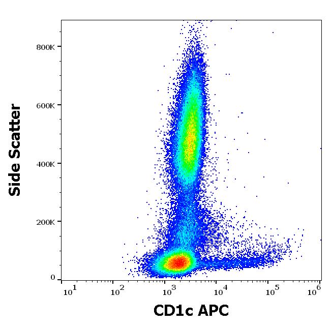
ARG42328 anti-CD1c antibody [L161] (APC) FACS image
Flow Cytometry: Human peripheral whole blood stained with ARG42328 anti-CD1c antibody [L161] (APC) at 10 µl / 100 µl of peripheral whole blood.
-
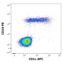
ARG42328 anti-CD1c antibody [L161] (APC) FACS image
Flow Cytometry: Human lymphocytes stained with ARG42328 anti-CD1c antibody [L161] (APC) at 10 µl / 100 µl of peripheral whole blood and ARG53783 anti-CD19 antibody [LT19] (PE) at 20 µl / 100 µl of peripheral whole blood.
-

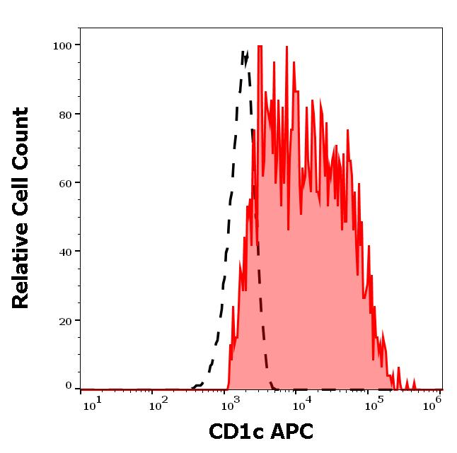
ARG42328 anti-CD1c antibody [L161] (APC) FACS image
Flow Cytometry: Separation of Human CD1c positive CD19 positive B cells (red-filled) from Human CD1c negative CD19 negative lymphocytes (black-dashed). Human peripheral whole blood stained with ARG42328 anti-CD1c antibody [L161] (APC) at 10 µl / 100 µl of peripheral whole blood.