ARG65538
anti-CD1b antibody [SN13] (FITC)
anti-CD1b antibody [SN13] (FITC) for Flow cytometry and Human
Immune System antibody
Overview
| Product Description | FITC-conjugated Mouse Monoclonal antibody [SN13] recognizes CD1b |
|---|---|
| Tested Reactivity | Hu |
| Tested Application | FACS |
| Specificity | The clone SN13 (also known as K5-1B8) recognizes CD1b, a 44 kDa type I glycoprotein associated with beta2-microglobulin. It is expressed on dendritic cells, Langerhans cells, thymocytes, and T acute lymphoblastic leukemia cells. |
| Host | Mouse |
| Clonality | Monoclonal |
| Clone | SN13 |
| Isotype | IgG1 |
| Target Name | CD1b |
| Antigen Species | Human |
| Immunogen | A cell membrane antigen preparation that was isolated from normal human thymocytes |
| Conjugation | FITC |
| Alternate Names | T-cell surface glycoprotein CD1b; CD1A; R1; CD antigen CD1b; CD1 |
Application Instructions
| Application Suggestion |
|
||||
|---|---|---|---|---|---|
| Application Note | * The dilutions indicate recommended starting dilutions and the optimal dilutions or concentrations should be determined by the scientist. |
Properties
| Form | Liquid |
|---|---|
| Purification Note | The purified antibody is conjugated with Fluorescein isothiocyanate (FITC) under optimum conditions. The reagent is free of unconjugated FITC and adjusted for direct use. No reconstitution is necessary. |
| Buffer | PBS, 15 mM Sodium azide and 0.2% (w/v) high-grade protease free BSA |
| Preservative | 15 mM Sodium azide |
| Stabilizer | 0.2% (w/v) high-grade protease free BSA |
| Storage Instruction | Aliquot and store in the dark at 2-8°C. Keep protected from prolonged exposure to light. Avoid repeated freeze/thaw cycles. Suggest spin the vial prior to opening. The antibody solution should be gently mixed before use. |
| Note | For laboratory research only, not for drug, diagnostic or other use. |
Bioinformation
| Database Links | |
|---|---|
| Gene Symbol | CD1B |
| Gene Full Name | CD1b molecule |
| Background | CD1b (also known as R1) together with CD1a and c, belongs to group 1 of CD1 antigens. These non-classical MHC-like glycoproteins serve as antigen-presenting molecules for a subset of T cells that responds to specific lipids and glycolipids found in the cell walls of bacterial pathogens or self-glycolipid antigens such as gangliosides, and they have also roles in antiviral immunity. The trafficking routes of the particular CD1 types differ and correspond to their ability to bind and present different groups of antigens. Besides non-peptide glycolipid antigen presentation to CD1-restricted T cells, CD1b has been implicated in thymocyte development. |
| Function | Antigen-presenting protein that binds self and non-self lipid and glycolipid antigens and presents them to T-cell receptors on natural killer T-cells. [UniProt] |
| Research Area | Immune System antibody |
| Calculated MW | 37 kDa |
Images (1) Click the Picture to Zoom In
-
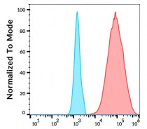
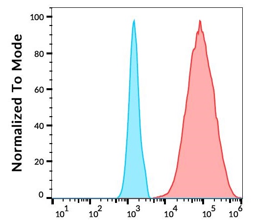
ARG65538 anti-CD1b antibody [SN13] (FITC) FACS image
Flow Cytometry: Separation of human CD1b positive dendritic cells differentiated upon monocyte stimulation (GM-CSF + IL-4) (red) from CD1b negative monocytes (blue). Stimulated (GM-CSF + IL-4) human peripheral blood mononuclear cells stained with ARG65538 anti-CD1b antibody [SN13] (FITC) (4 µl reagent / 10^6 cells in 100 µl of cell suspension).
Clone References
Id1 and NF-κB promote the generation of CD133+ and BMI-1+ keratinocytes and the growth of xenograft tumors in mice.
WB / Human


Influence of Mycobacterium bovis bacillus Calmette Guérin on in vitro induction of CD1 molecules in human adherent mononuclear cells.


