ARG43274
anti-CD163 antibody
anti-CD163 antibody for Flow cytometry,IHC-Formalin-fixed paraffin-embedded sections,Western blot and Human,Mouse,Rat
Overview
| Product Description | Rabbit Polyclonal antibody recognizes CD163 |
|---|---|
| Tested Reactivity | Hu, Ms, Rat |
| Tested Application | FACS, IHC-P, WB |
| Host | Rabbit |
| Clonality | Polyclonal |
| Isotype | IgG |
| Target Name | CD163 |
| Antigen Species | Human |
| Immunogen | Recombinant protein corresponding to T47-E201 of Human CD163. |
| Conjugation | Un-conjugated |
| Alternate Names | sCD163; M130; Scavenger receptor cysteine-rich type 1 protein M130; MM130; CD antigen CD163; Hemoglobin scavenger receptor |
Application Instructions
| Application Suggestion |
|
||||||||
|---|---|---|---|---|---|---|---|---|---|
| Application Note | IHC-P: Antigen Retrieval: Heat mediation was performed in EDTA buffer (pH 8.0). * The dilutions indicate recommended starting dilutions and the optimal dilutions or concentrations should be determined by the scientist. |
||||||||
| Positive Control | HeLa, Human placenta and NIH/3T3 | ||||||||
| Observed Size | ~ 140 kDa |
Properties
| Form | Liquid |
|---|---|
| Purification | Affinity purification with immunogen. |
| Buffer | 0.2% Na2HPO4, 0.9% NaCl, 0.05% Sodium azide and 4% Trehalose. |
| Preservative | 0.05% Sodium azide |
| Stabilizer | 4% Trehalose |
| Concentration | 0.5 mg/ml |
| Storage Instruction | For continuous use, store undiluted antibody at 2-8°C for up to a week. For long-term storage, aliquot and store at -20°C or below. Storage in frost free freezers is not recommended. Avoid repeated freeze/thaw cycles. Suggest spin the vial prior to opening. The antibody solution should be gently mixed before use. |
| Note | For laboratory research only, not for drug, diagnostic or other use. |
Bioinformation
| Database Links |
Swiss-port # Q2VLH6 Mouse Scavenger receptor cysteine-rich type 1 protein M130 Swiss-port # Q86VB7 Human Scavenger receptor cysteine-rich type 1 protein M130 |
|---|---|
| Gene Symbol | CD163 |
| Gene Full Name | CD163 molecule |
| Background | The protein encoded by this gene is a member of the scavenger receptor cysteine-rich (SRCR) superfamily, and is exclusively expressed in monocytes and macrophages. It functions as an acute phase-regulated receptor involved in the clearance and endocytosis of hemoglobin/haptoglobin complexes by macrophages, and may thereby protect tissues from free hemoglobin-mediated oxidative damage. This protein may also function as an innate immune sensor for bacteria and inducer of local inflammation. Alternatively spliced transcript variants encoding different isoforms have been described for this gene. [provided by RefSeq, Aug 2011] |
| Function | Acute phase-regulated receptor involved in clearance and endocytosis of hemoglobin/haptoglobin complexes by macrophages and may thereby protect tissues from free hemoglobin-mediated oxidative damage. May play a role in the uptake and recycling of iron, via endocytosis of hemoglobin/haptoglobin and subsequent breakdown of heme. Binds hemoglobin/haptoglobin complexes in a calcium-dependent and pH-dependent manner. Exhibits a higher affinity for complexes of hemoglobin and multimeric haptoglobin of HP*1F phenotype than for complexes of hemoglobin and dimeric haptoglobin of HP*1S phenotype. Induces a cascade of intracellular signals that involves tyrosine kinase-dependent calcium mobilization, inositol triphosphate production and secretion of IL6 and CSF1. Isoform 3 exhibits the higher capacity for ligand endocytosis and the more pronounced surface expression when expressed in cells. After shedding, the soluble form (sCD163) may play an anti-inflammatory role, and may be a valuable diagnostic parameter for monitoring macrophage activation in inflammatory conditions. [UniProt] |
| Cellular Localization | Soluble CD163: Secreted. Cell membrane; Single-pass type I membrane protein. Note=Isoform 1 and isoform 2 show a lower surface expression when expressed in cells. [UniProt] |
| Highlight | Related products: CD163 antibodies; CD163 ELISA Kits; CD163 Duos / Panels; Anti-Rabbit IgG secondary antibodies; Related news: RIP1 activation and pathogenesis of NASH |
| Calculated MW | 125 kDa |
| PTM | A soluble form (sCD163) is produced by proteolytic shedding which can be induced by lipopolysaccharide, phorbol ester and Fc region of immunoglobulin gamma. This cleavage is dependent on protein kinase C and tyrosine kinases and can be blocked by protease inhibitors. The shedding is inhibited by the tissue inhibitor of metalloproteinase TIMP3, and thus probably induced by membrane-bound metalloproteinases ADAMs. Phosphorylated. [UniProt] |
Images (7) Click the Picture to Zoom In
-
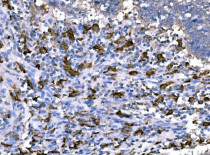
ARG43274 anti-CD163 antibody IHC-P image
Immunohistochemistry: Paraffin-embedded Human tonsil tissue. Antigen Retrieval: Heat mediation was performed in EDTA buffer (pH 8.0). The tissue section was blocked with 10% goat serum. The tissue section was then stained with ARG43274 anti-CD163 antibody at 1 µg/ml dilution, overnight at 4°C.
-

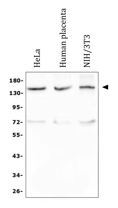
ARG43274 anti-CD163 antibody WB image
Western blot: 50 µg of sample under reducing conditions. HeLa, Human placenta and NIH/3T3 whole cell lysates stained with ARG43274 anti-CD163 antibody at 0.5 µg/ml dilution, overnight at 4°C.
-

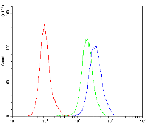
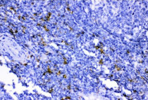
ARG43274 anti-CD163 antibody FACS image
Flow Cytometry: THP-1 cells were blocked with 10% normal goat serum and then stained with ARG43274 anti-CD163 antibody (blue) at 1 µg/10^6 cells for 30 min at 20°C, followed by incubation with DyLight®488 labelled secondary antibody. Isotype control antibody (green) was rabbit IgG (1 µg/10^6 cells) used under the same conditions. Unlabelled sample (red) was also used as a control.
-
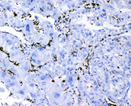
ARG43274 anti-CD163 antibody IHC-P image
Immunohistochemistry: Paraffin-embedded Human liver cancer tissue. Antigen Retrieval: Heat mediation was performed in EDTA buffer (pH 8.0). The tissue section was blocked with 10% goat serum. The tissue section was then stained with ARG43274 anti-CD163 antibody at 1 µg/ml dilution, overnight at 4°C.
-
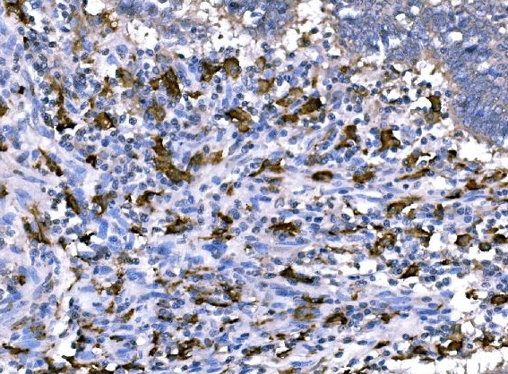
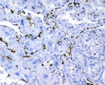
ARG43274 anti-CD163 antibody IHC-P image
Immunohistochemistry: Paraffin-embedded Human rectal cancer tissue. Antigen Retrieval: Heat mediation was performed in EDTA buffer (pH 8.0). The tissue section was blocked with 10% goat serum. The tissue section was then stained with ARG43274 anti-CD163 antibody at 1 µg/ml dilution, overnight at 4°C.
-
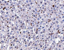
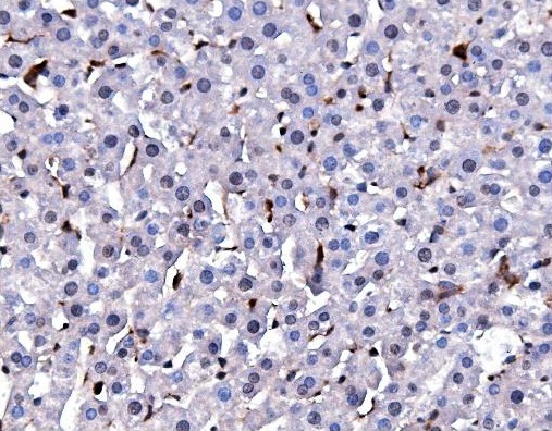
ARG43274 anti-CD163 antibody IHC-P image
Immunohistochemistry: Paraffin-embedded Rat liver tissue. Antigen Retrieval: Heat mediation was performed in EDTA buffer (pH 8.0). The tissue section was blocked with 10% goat serum. The tissue section was then stained with ARG43274 anti-CD163 antibody at 1 µg/ml dilution, overnight at 4°C.
-
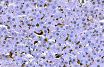
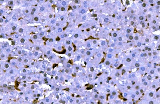
ARG43274 anti-CD163 antibody IHC-P image
Immunohistochemistry: Paraffin-embedded Rat liver tissue. Antigen Retrieval: Heat mediation was performed in EDTA buffer (pH 8.0). The tissue section was blocked with 10% goat serum. The tissue section was then stained with ARG43274 anti-CD163 antibody at 1 µg/ml dilution, overnight at 4°C.