ARG22980
anti-CD16 antibody [G7] (PE)
anti-CD16 antibody [G7] (PE) for Flow cytometry and Pig
Developmental Biology antibody; Immune System antibody; General Lymphocyte Marker Study antibody; Natural killer cells antibody
Overview
| Product Description | PE-conjugated Mouse Monoclonal antibody [G7] recognizes CD16 Mouse anti Pig CD16, clone G7 recognizes porcine CD16 also known as Fc-gamma RIII or the low affinity IgG (Fc) receptor III. Clone G7 was clustered as CD16 at the Second International Workshop to Define Swine Cluster of Differentiation (CD) Antigens (Saalmuller et al. 1998).Mouse anti pig CD16 immunoprecipitates a protein of ~40 kDa from porcine neutrophils and NK cells (Wierda et al. 1993). Subsequent cloning and characterization of the G7 molecule indicated that G7 was the porcine homologue of Human CD16 (Halloran et al. 1994). |
|---|---|
| Tested Reactivity | Pig |
| Tested Application | FACS |
| Host | Mouse |
| Clonality | Monoclonal |
| Clone | G7 |
| Isotype | IgG1 |
| Target Name | CD16 |
| Antigen Species | Pig |
| Immunogen | Porcine peripheral blood leucocytes |
| Conjugation | PE |
| Alternate Names | FCRIIIA; FcRIIIa; CD antigen CD16a; Fc-gamma RIII-alpha; FCR-10; FcR-10; FCRIII; FCG3; Low affinity immunoglobulin gamma Fc region receptor III-A; FCGRIII; CD16; Fc-gamma RIIIa; IgG Fc receptor III-2; IMD20; CD16A; IGFR3; CD16a antigen; FCGR3; FcRIII; Fc-gamma RIII |
Application Instructions
| Application Suggestion |
|
||||
|---|---|---|---|---|---|
| Application Note | FACS: Use 10 µl of the suggested working dilution to label 10^6 cells in 100 µl. * The dilutions indicate recommended starting dilutions and the optimal dilutions or concentrations should be determined by the scientist. |
Properties
| Form | Liquid |
|---|---|
| Purification | Purification with Protein A. |
| Buffer | PBS, 0.09% Sodium azide, 1% BSA and 5% Sucrose |
| Preservative | 0.09% Sodium azide |
| Stabilizer | 1% BSA and 5% Sucrose |
| Storage Instruction | Aliquot and store in the dark at 2-8°C. Keep protected from prolonged exposure to light. Avoid repeated freeze/thaw cycles. Suggest spin the vial prior to opening. The antibody solution should be gently mixed before use. |
| Note | For laboratory research only, not for drug, diagnostic or other use. |
Bioinformation
| Gene Symbol | FCGR3A |
|---|---|
| Gene Full Name | Fc fragment of IgG receptor IIIa |
| Background | This gene encodes a receptor for the Fc portion of immunoglobulin G, and it is involved in the removal of antigen-antibody complexes from the circulation, as well as other other antibody-dependent responses. This gene (FCGR3A) is highly similar to another nearby gene (FCGR3B) located on chromosome 1. The receptor encoded by this gene is expressed on natural killer (NK) cells as an integral membrane glycoprotein anchored through a transmembrane peptide, whereas FCGR3B is expressed on polymorphonuclear neutrophils (PMN) where the receptor is anchored through a phosphatidylinositol (PI) linkage. Mutations in this gene have been linked to susceptibility to recurrent viral infections, susceptibility to systemic lupus erythematosus, and alloimmune neonatal neutropenia. Alternatively spliced transcript variants encoding different isoforms have been found for this gene. [provided by RefSeq, Jul 2008] |
| Function | Receptor for the Fc region of IgG. Binds complexed or aggregated IgG and also monomeric IgG. Mediates antibody-dependent cellular cytotoxicity (ADCC) and other antibody-dependent responses, such as phagocytosis. [UniProt] |
| Highlight | Related products: CD16 antibodies; CD16 ELISA Kits; CD16 Duos / Panels; Anti-Mouse IgG secondary antibodies; Related news: Tumor-Infiltrating Lymphocytes (TILs) |
| Research Area | Developmental Biology antibody; Immune System antibody; General Lymphocyte Marker Study antibody; Natural killer cells antibody |
| Calculated MW | 29 kDa |
| PTM | Glycosylated. Contains high mannose- and complex-type oligosaccharides. Glycosylation at Asn-180 is mandatory for high affinity binding to the Fc and for discrimination between fucosylated and afucosylated IgG glycoforms. The soluble form is produced by a proteolytic cleavage. |
Images (3) Click the Picture to Zoom In
-
ARG22980 anti-CD16 antibody [G7] (PE) FACS image
Flow Cytometry: Porcine peripheral blood granulocytes stained with ARG22980 anti-CD16 antibody [G7] (PE).
-
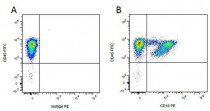
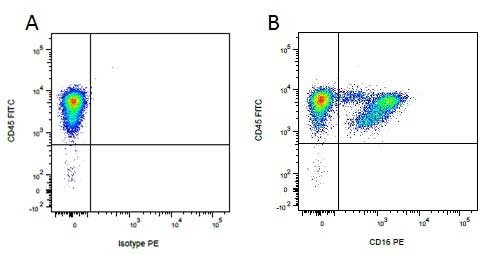
ARG22980 anti-CD16 antibody [G7] (PE) FACS image
Flow Cytometry: Figure A. FITC conjugated mouse anti porcine CD45 and PE conjugated mouse IgG1 isotype control. Figure B. FITC conjugated mouse anti porcine CD45 and ARG22980 anti-CD16 antibody [G7] (PE). All experiments performed on red cell lysed porcine blood gated on mononuclear cells.
-
ARG22980 anti-CD16 antibody [G7] (PE) FACS image
Flow Cytometry: Figure A. ARG22980 anti-CD16 antibody [G7] (PE) and FITC conjugated mouse IgG1 isotype control. Figure B. ARG22980 anti-CD16 antibody [G7] (PE) and FITC conjugated mouse anti porcine CD45. All experiments performed on red cell lysed porcine blood gated on mononuclear cells.
Specific References
The antibodies against the A137R protein drive antibody-dependent enhancement of African swine fever virus infection in porcine alveolar macrophages
Blocking / Pig


