ARG40693
anti-CD16 antibody
anti-CD16 antibody for Flow cytometry,ICC/IF,IHC-Formalin-fixed paraffin-embedded sections and Human
Developmental Biology antibody; Immune System antibody; General Lymphocyte Marker Study antibody; Natural killer cells antibody
Overview
| Product Description | Rabbit Polyclonal antibody recognizes CD16 |
|---|---|
| Tested Reactivity | Hu |
| Tested Application | FACS, ICC/IF, IHC-P |
| Host | Rabbit |
| Clonality | Polyclonal |
| Isotype | IgG |
| Target Name | CD16 |
| Antigen Species | Human |
| Immunogen | Recombinant protein corresponding to Q101-D166 of Human CD16. |
| Conjugation | Un-conjugated |
| Alternate Names | FCRIIIA; FcRIIIa; CD antigen CD16a; Fc-gamma RIII-alpha; FCR-10; FcR-10; FCRIII; FCG3; Low affinity immunoglobulin gamma Fc region receptor III-A; FCGRIII; CD16; Fc-gamma RIIIa; IgG Fc receptor III-2; IMD20; CD16A; IGFR3; CD16a antigen; FCGR3; FcRIII; Fc-gamma RIII |
Application Instructions
| Application Suggestion |
|
||||||||
|---|---|---|---|---|---|---|---|---|---|
| Application Note | IHC-P: Antigen Retrieval: Heat mediation was performed in EDTA buffer (pH 8.0). * The dilutions indicate recommended starting dilutions and the optimal dilutions or concentrations should be determined by the scientist. |
Properties
| Form | Liquid |
|---|---|
| Buffer | 0.2% Na2HPO4, 0.9% NaCl, 0.05% Sodium azide and 4% Trehalose. |
| Preservative | 0.05% Sodium azide |
| Stabilizer | 4% Trehalose |
| Concentration | 0.5 mg/ml |
| Storage Instruction | For continuous use, store undiluted antibody at 2-8°C for up to a week. For long-term storage, aliquot and store at -20°C or below. Storage in frost free freezers is not recommended. Avoid repeated freeze/thaw cycles. Suggest spin the vial prior to opening. The antibody solution should be gently mixed before use. |
| Note | For laboratory research only, not for drug, diagnostic or other use. |
Bioinformation
| Database Links |
Swiss-port # P08637 Human Low affinity immunoglobulin gamma Fc region receptor III-A |
|---|---|
| Gene Symbol | FCGR3A |
| Gene Full Name | Fc fragment of IgG, low affinity IIIa, receptor (CD16a) |
| Background | This gene encodes a receptor for the Fc portion of immunoglobulin G, and it is involved in the removal of antigen-antibody complexes from the circulation, as well as other other antibody-dependent responses. This gene (FCGR3A) is highly similar to another nearby gene (FCGR3B) located on chromosome 1. The receptor encoded by this gene is expressed on natural killer (NK) cells as an integral membrane glycoprotein anchored through a transmembrane peptide, whereas FCGR3B is expressed on polymorphonuclear neutrophils (PMN) where the receptor is anchored through a phosphatidylinositol (PI) linkage. Mutations in this gene have been linked to susceptibility to recurrent viral infections, susceptibility to systemic lupus erythematosus, and alloimmune neonatal neutropenia. Alternatively spliced transcript variants encoding different isoforms have been found for this gene. [provided by RefSeq, Jul 2008] |
| Function | Receptor for the Fc region of IgG. Binds complexed or aggregated IgG and also monomeric IgG. Mediates antibody-dependent cellular cytotoxicity (ADCC) and other antibody-dependent responses, such as phagocytosis. [UniProt] |
| Cellular Localization | Cell membrane; Single-pass type I membrane protein. Secreted. Note=Exists also as a soluble receptor. [UniProt] |
| Highlight | Related products: CD16 antibodies; CD16 ELISA Kits; CD16 Duos / Panels; Anti-Rabbit IgG secondary antibodies; Related news: Tumor-Infiltrating Lymphocytes (TILs) |
| Research Area | Developmental Biology antibody; Immune System antibody; General Lymphocyte Marker Study antibody; Natural killer cells antibody |
| Calculated MW | 29 kDa |
| PTM | Glycosylated. Contains high mannose- and complex-type oligosaccharides. Glycosylation at Asn-180 is mandatory for high affinity binding to the Fc and for discrimination between fucosylated and afucosylated IgG glycoforms. The soluble form is produced by a proteolytic cleavage. [UniProt] |
Images (5) Click the Picture to Zoom In
-

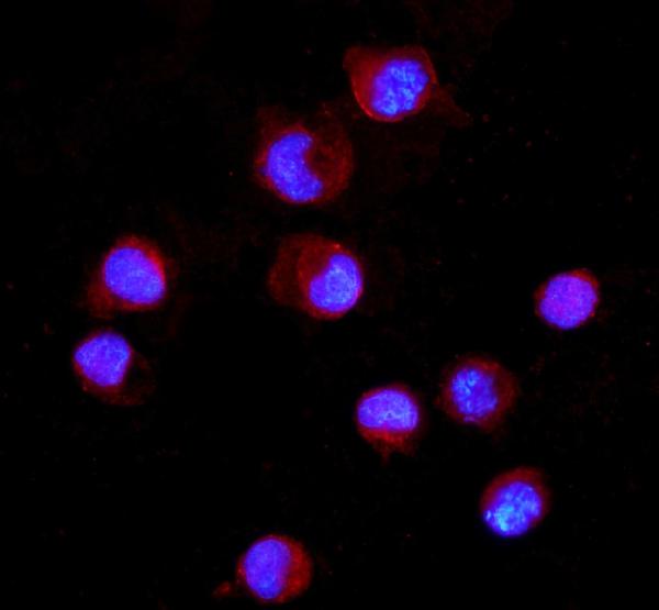
ARG40693 anti-CD16 antibody ICC/IF image
Immunofluorescence: K562 cells were blocked with 10% goat serum and then stained with ARG40693 anti-CD16 antibody (red) at 5 µg/ml dilution, overnight at 4°C. DAPI (blue) for nuclear staining.
-
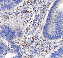
ARG40693 anti-CD16 antibody IHC-P image
Immunohistochemistry: Paraffin-embedded Human liver cancer tissue. Antigen Retrieval: Heat mediation was performed in EDTA buffer (pH 8.0). The tissue section was blocked with 10% goat serum. The tissue section was then stained with ARG40693 anti-CD16 antibody at 2 µg/ml dilution, overnight at 4°C.
-
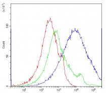
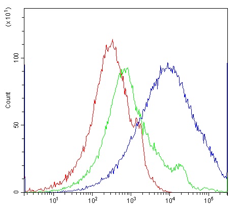
ARG40693 anti-CD16 antibody FACS image
Flow Cytometry: Human PBMC cells were blocked with 10% normal goat serum and then stained with ARG40693 anti-CD16 antibody (blue) at 1 µg/10^6 cells for 30 min at 20°C, followed by incubation with DyLight®488 labelled secondary antibody. Isotype control antibody (green) was rabbit IgG (1 µg/10^6 cells) used under the same conditions. Unlabelled sample (red) was also used as a control.
-
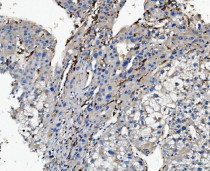
ARG40693 anti-CD16 antibody IHC-P image
Immunohistochemistry: Paraffin-embedded Human rectal cancer tissue. Antigen Retrieval: Heat mediation was performed in EDTA buffer (pH 8.0). The tissue section was blocked with 10% goat serum. The tissue section was then stained with ARG40693 anti-CD16 antibody at 2 µg/ml dilution, overnight at 4°C.
-
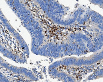
ARG40693 anti-CD16 antibody IHC-P image
Immunohistochemistry: Paraffin-embedded Human rectal cancer tissue. Antigen Retrieval: Heat mediation was performed in EDTA buffer (pH 8.0). The tissue section was blocked with 10% goat serum. The tissue section was then stained with ARG40693 anti-CD16 antibody at 2 µg/ml dilution, overnight at 4°C.