ARG44057
anti-CD158a + CD158g CD158h antibody [HP-MA4] (PE-Cyanine 7)
anti-CD158a + CD158g CD158h antibody [HP-MA4] (PE-Cyanine 7) for Flow cytometry and Human
Overview
| Product Description | PE-Cyanine 7-conjugated Mouse Monoclonal antibody recognizes CD158a + CD158g CD158h |
|---|---|
| Tested Reactivity | Hu |
| Tested Application | FACS |
| Specificity | The mouse monoclonal antibody HP-MA4 recognizes an extracellular epitope of CD158 isoforms KIR2DL1 (CD158a), KIR2DS5 (CD158g), KIR2DS1 (CD158h), and KIR2DS3. It does not recognize the isoforms CD158b1,d,f,i,j. |
| Host | Mouse |
| Clonality | Monoclonal |
| Clone | HP-MA4 |
| Isotype | IgG2a |
| Target Name | CD158a + CD158g+ CD158h |
| Antigen Species | Human |
| Immunogen | Human NK cell line LB2 |
| Conjugation | PE-Cyanine 7 |
| Alternate Names | KIR2DL1; Killer Cell Immunoglobulin Like Receptor, Two Ig Domains And Long Cytoplasmic Tail 1; CD158A ; KIR2DS5; Killer Cell Immunoglobulin Like Receptor, Two Ig Domains And Short Cytoplasmic Tail 5; CD158G; KIR2DS1; Killer Cell Immunoglobulin Like Receptor, Two Ig Domains And Short Cytoplasmic Tail 1; CD158H |
Application Instructions
| Application Suggestion |
|
||||
|---|---|---|---|---|---|
| Application Note | * The dilutions indicate recommended starting dilutions and the optimal dilutions or concentrations should be determined by the scientist. |
Properties
| Form | Liquid |
|---|---|
| Buffer | PBS (pH 7.4) and 15 mM Sodium azide |
| Preservative | 15 mM Sodium azide |
| Storage Instruction | Aliquot and store in the dark at 2-8°C. Keep protected from prolonged exposure to light. Avoid repeated freeze/thaw cycles. Suggest spin the vial prior to opening. The antibody solution should be gently mixed before use. |
| Note | For laboratory research only, not for drug, diagnostic or other use. |
Bioinformation
| Gene Symbol | KIR2DL1; KIR2DS5; KIR2DS1 |
|---|---|
| Gene Full Name | Killer Cell Immunoglobulin Like Receptor, Two Ig Domains And Long Cytoplasmic Tail 1; Killer Cell Immunoglobulin Like Receptor, Two Ig Domains And Short Cytoplasmic Tail 5; Killer Cell Immunoglobulin Like Receptor, Two Ig Domains And Short Cytoplasmic Tail 1 |
| Background | Killer cell immunoglobulin-like receptors (KIRs) are transmembrane glycoproteins expressed by natural killer cells and subsets of T cells. The KIR genes are polymorphic and highly homologous and they are found in a cluster on chromosome 19q13.4 within the 1 Mb leukocyte receptor complex (LRC). The gene content of the KIR gene cluster varies among haplotypes, although several "framework" genes are found in all haplotypes (KIR3DL3, KIR3DP1, KIR3DL4, KIR3DL2). The KIR proteins are classified by the number of extracellular immunoglobulin domains (2D or 3D) and by whether they have a long (L) or short (S) cytoplasmic domain. KIR proteins with the long cytoplasmic domain transduce inhibitory signals upon ligand binding via an immune tyrosine-based inhibitory motif (ITIM), while KIR proteins with the short cytoplasmic domain lack the ITIM motif and instead associate with the TYRO protein tyrosine kinase binding protein to transduce activating signals. The ligands for several KIR proteins are subsets of HLA class I molecules; thus, KIR proteins are thought to play an important role in regulation of the immune response. Killer cell immunoglobulin-like receptors (KIRs) are transmembrane glycoproteins expressed by natural killer cells and subsets of T cells. The KIR genes are polymorphic and highly homologous and they are found in a cluster on chromosome 19q13.4 within the 1 Mb leukocyte receptor complex (LRC). The gene content of the KIR gene cluster varies among haplotypes, although several "framework" genes are found in all haplotypes (KIR3DL3, KIR3DP1, KIR3DL4, KIR3DL2). The KIR proteins are classified by the number of extracellular immunoglobulin domains (2D or 3D) and by whether they have a long (L) or short (S) cytoplasmic domain. KIR proteins with the long cytoplasmic domain transduce inhibitory signals upon ligand binding via an immune tyrosine-based inhibitory motif (ITIM), while KIR proteins with the short cytoplasmic domain lack the ITIM motif and instead associate with the TYRO protein tyrosine kinase binding protein to transduce activating signals. The ligands for several KIR proteins are subsets of HLA class I molecules; thus, KIR proteins are thought to play an important role in regulation of the immune response. Killer cell immunoglobulin-like receptors (KIRs) are transmembrane glycoproteins expressed by natural killer cells and subsets of T cells. The KIR genes are polymorphic and highly homologous and they are found in a cluster on chromosome 19q13.4 within the 1 Mb leukocyte receptor complex (LRC). The gene content of the KIR gene cluster varies among haplotypes, although several "framework" genes are found in all haplotypes (KIR3DL3, KIR3DP1, KIR3DL4, KIR3DL2). The KIR proteins are classified by the number of extracellular immunoglobulin domains (2D or 3D) and by whether they have a long (L) or short (S) cytoplasmic domain. KIR proteins with the long cytoplasmic domain transduce inhibitory signals upon ligand binding via an immune tyrosine-based inhibitory motif (ITIM), while KIR proteins with the short cytoplasmic domain lack the ITIM motif and instead associate with the TYRO protein tyrosine kinase binding protein to transduce activating signals. The ligands for several KIR proteins are subsets of HLA class I molecules; thus, KIR proteins are thought to play an important role in regulation of the immune response. |
| Function | Receptor on natural killer (NK) cells for some HLA-C alleles such as w4 and w6. Inhibits the activity of NK cells thus preventing cell lysis. Receptor functions are attenuated even lost in some alleles, such as KIR2DS5*002 reprensented in this entry. Receptor on natural killer (NK) cells for some HLA-C alleles such as w6. Does not inhibit the activity of NK cells. |
| Cellular Localization | Cell membrane, Membrane |
| PTM | Disulfide bond, Glycoprotein |
Images (1) Click the Picture to Zoom In
-
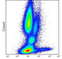
ARG44057 anti-CD158a + CD158g CD158h antibody [HP-MA4] (PE-Cyanine 7) FACS image
Flow Cytometry: Human peripheral whole blood stained with ARG44057 anti-CD158a + CD158g CD158h antibody [HP-MA4] (PE-Cyanine 7) at 10 μl / 10^6 cells dilution.


