ARG45400
anti-CD148 / DEP1 antibody
anti-CD148 / DEP1 antibody for Flow cytometry,IHC-Formalin-fixed paraffin-embedded sections,Western blot and Human,Rat
Overview
| Product Description | Rabbit Polyclonal antibody recognizes CD148 / DEP1 |
|---|---|
| Tested Reactivity | Hu, Rat |
| Tested Application | FACS, IHC-P, WB |
| Host | Rabbit |
| Clonality | Polyclonal |
| Isotype | IgG |
| Target Name | CD148 / DEP1 |
| Antigen Species | Human |
| Immunogen | Recombinant protein containing to human CD148 / DEP1. |
| Conjugation | Un-conjugated |
| Alternate Names | R-PTP-J; Density-enhanced phosphatase 1; R-PTP-ETA; DEP-1; SCC1; DEP1; CD antigen CD148; Receptor-type tyrosine-protein phosphatase eta; Protein-tyrosine phosphatase eta; Protein-tyrosine phosphatase receptor type J; EC 3.1.3.48; HPTPeta; HPTP eta; R-PTP-eta; CD148 |
Application Instructions
| Application Suggestion |
|
||||||||
|---|---|---|---|---|---|---|---|---|---|
| Application Note | * The dilutions indicate recommended starting dilutions and the optimal dilutions or concentrations should be determined by the scientist. | ||||||||
| Observed Size | 240 kDa |
Properties
| Form | Liquid |
|---|---|
| Purification | Affinity purified |
| Buffer | 0.2% Na2HPO4, 0.9% NaCl and 4% Trehalose. |
| Stabilizer | 4% Trehalose |
| Concentration | 0.5 mg/ml |
| Storage Instruction | For continuous use, store undiluted antibody at 2-8°C for up to a week. For long-term storage, aliquot and store at -20°C or below. Storage in frost free freezers is not recommended. Avoid repeated freeze/thaw cycles. Suggest spin the vial prior to opening. The antibody solution should be gently mixed before use. |
| Note | For laboratory research only, not for drug, diagnostic or other use. |
Bioinformation
| Database Links |
Swiss-port # Q12913 Human Receptor-type tyrosine-protein phosphatase eta |
|---|---|
| Gene Symbol | PTPRJ |
| Gene Full Name | protein tyrosine phosphatase, receptor type, J |
| Background | The protein encoded by this gene is a member of the protein tyrosine phosphatase (PTP) family. PTPs are known to be signaling molecules that regulate a variety of cellular processes, including cell growth, differentiation, mitotic cycle, and oncogenic transformation. This PTP possesses an extracellular region containing five fibronectin type III repeats, a single transmembrane region, and a single intracytoplasmic catalytic domain, and thus represents a receptor-type PTP. This protein is present in all hematopoietic lineages, and was shown to negatively regulate T cell receptor signaling possibly through interfering with the phosphorylation of Phospholipase C Gamma 1 and Linker for Activation of T Cells. This protein can also dephosphorylate the PDGF beta receptor, and may be involved in UV-induced signal transduction. Multiple transcript variants encoding different isoforms have been found for this gene. [provided by RefSeq, Jul 2008] |
| Function | Tyrosine phosphatase which dephosphorylates or contributes to the dephosphorylation of CTNND1, FLT3, PDGFRB, MET, RET (variant MEN2A), KDR, LYN, SRC, MAPK1, MAPK3, EGFR, TJP1, OCLN, PIK3R1 and PIK3R2. Plays a role in cell adhesion, migration, proliferation and differentiation. Involved in vascular development. Regulator of macrophage adhesion and spreading. Positively affects cell-matrix adhesion. Positive regulator of platelet activation and thrombosis. Negative regulator of cell proliferation. Negative regulator of PDGF-stimulated cell migration; through dephosphorylation of PDGFR. Positive regulator of endothelial cell survival, as well as of VEGF-induced SRC and AKT activation; through KDR dephosphorylation. Negative regulator of EGFR signaling pathway; through EGFR dephosphorylation. Enhances the barrier function of epithelial junctions during reassembly. Negatively regulates T-cell receptor (TCR) signaling. Upon T-cell TCR activation, it is up-regulated and excluded from the immunological synapses, while upon T-cell-antigen presenting cells (APC) disengagement, it is no longer excluded and can dephosphorylate PLCG1 and LAT to down-regulate prolongation of signaling. [UniProt] |
| Cellular Localization | Cell junction; Cell membrane; Cell projection; Membrane; Secreted. [UniProt] |
| Calculated MW | 146 kDa |
| PTM | N- and O-glycosylated. [UniProt] |
Images (5) Click the Picture to Zoom In
-

ARG45400 anti-CD148 / DEP1 antibody IHC-P image
Immunohistochemistry: Human rectal cancer stained with ARG45400 anti-CD148 / DEP1 antibody at 2 μg/ml dilution.
-
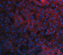
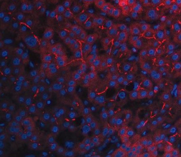
ARG45400 anti-CD148 / DEP1 antibody ICC/IF image
Immunofluorescence: Human liver cancer stained with ARG45400 anti-CD148 / DEP1 antibody at 5 μg/ml dilution.
-
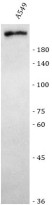
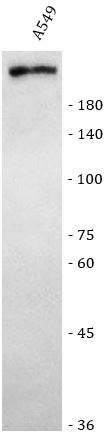
ARG45400 anti-CD148 / DEP1 antibody WB image
Western blot: A549 stained with ARG45400 anti-CD148 / DEP1 antibody at 0.5 μg/ml dilution.
-
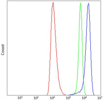
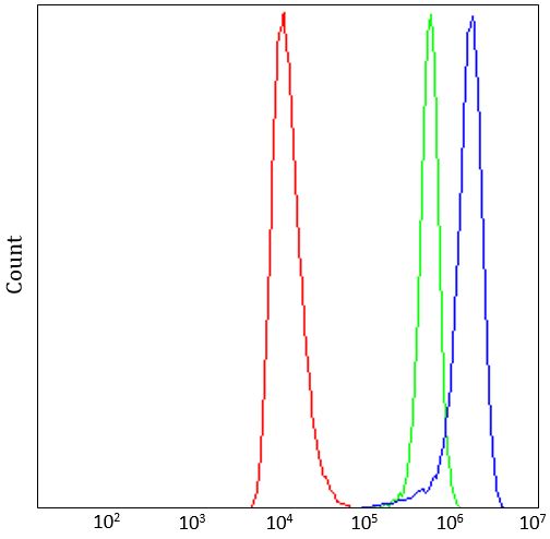
ARG45400 anti-CD148 / DEP1 antibody FACS image
Flow Cytometry: HEL stained with ARG45400 anti-CD148 / DEP1 antibody at 1 µg/10^6 cells dilution.
-
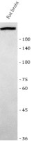
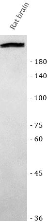
ARG45400 anti-CD148 / DEP1 antibody WB image
Western blot: Rat brain stained with ARG45400 anti-CD148 / DEP1 antibody at 0.5 μg/ml dilution.