ARG41554
anti-CD147 antibody
anti-CD147 antibody for Flow cytometry,ICC/IF,IHC-Formalin-fixed paraffin-embedded sections,Western blot and Human
Overview
| Product Description | Rabbit Polyclonal antibody recognizes CD147 |
|---|---|
| Tested Reactivity | Hu |
| Tested Application | FACS, ICC/IF, IHC-P, WB |
| Host | Rabbit |
| Clonality | Polyclonal |
| Isotype | IgG |
| Target Name | CD147 |
| Antigen Species | Human |
| Immunogen | Recombinant protein corresponding to E138-A323 of Human CD147. |
| Conjugation | Un-conjugated |
| Alternate Names | OK; Tumor cell-derived collagenase stimulatory factor; Leukocyte activation antigen M6; TCSF; Extracellular matrix metalloproteinase inducer; OK blood group antigen; CD147; Basigin; M6; EMMPRIN; Collagenase stimulatory factor; CD antigen CD147; 5F7 |
Application Instructions
| Application Suggestion |
|
||||||||||
|---|---|---|---|---|---|---|---|---|---|---|---|
| Application Note | IHC-P: Antigen Retrieval: Heat mediation was performed in Citrate buffer (pH 6.0, epitope retrieval solution) for 20 min. * The dilutions indicate recommended starting dilutions and the optimal dilutions or concentrations should be determined by the scientist. |
||||||||||
| Observed Size | ~ 35 kDa (nonglycosylated); ~ 50 kDa (glycosylated) |
Properties
| Form | Liquid |
|---|---|
| Purification | Affinity purification with immunogen. |
| Buffer | 0.2% Na2HPO4, 0.9% NaCl, 0.05% Sodium azide and 5% BSA. |
| Preservative | 0.05% Sodium azide |
| Stabilizer | 5% BSA |
| Concentration | 0.5 mg/ml |
| Storage Instruction | For continuous use, store undiluted antibody at 2-8°C for up to a week. For long-term storage, aliquot and store at -20°C or below. Storage in frost free freezers is not recommended. Avoid repeated freeze/thaw cycles. Suggest spin the vial prior to opening. The antibody solution should be gently mixed before use. |
| Note | For laboratory research only, not for drug, diagnostic or other use. |
Bioinformation
| Database Links | |
|---|---|
| Gene Symbol | BSG |
| Gene Full Name | basigin (Ok blood group) |
| Background | The protein encoded by this gene is a plasma membrane protein that is important in spermatogenesis, embryo implantation, neural network formation, and tumor progression. The encoded protein is also a member of the immunoglobulin superfamily. Multiple transcript variants encoding different isoforms have been found for this gene. [provided by RefSeq, Jul 2008] |
| Function | Plays an important role in targeting the monocarboxylate transporters SLC16A1, SLC16A3 and SLC16A8 to the plasma membrane. Plays pivotal roles in spermatogenesis, embryo implantation, neural network formation and tumor progression. Stimulates adjacent fibroblasts to produce matrix metalloproteinases (MMPS). Seems to be a receptor for oligomannosidic glycans. In vitro, promotes outgrowth of astrocytic processes. [UniProt] |
| Cellular Localization | Cell membrane; Single-pass type I membrane protein. Melanosome. Note=Identified by mass spectrometry in melanosome fractions from stage I to stage IV. In spermatozoa, localized on the principal piece of caput and in the middle piece during transit in the corpus and cauda epididymides (By similarity). [UniProt] |
| Calculated MW | 42 kDa |
| PTM | N-glycosylated. [UniProt] |
Images (8) Click the Picture to Zoom In
-
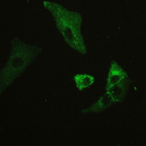
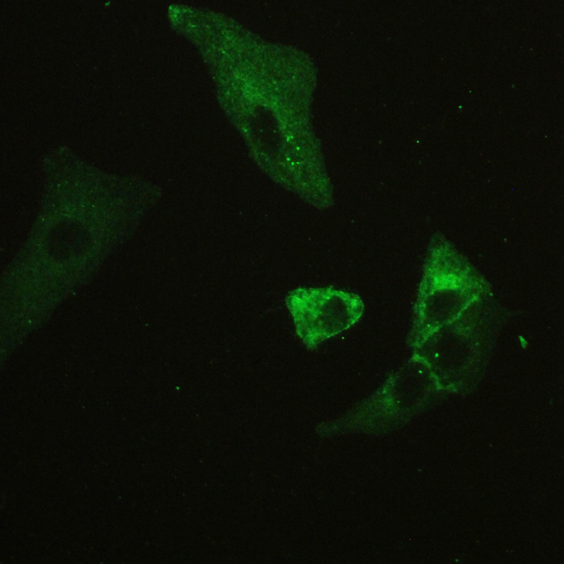
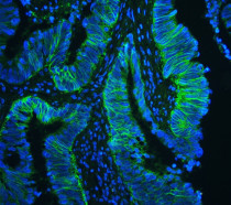
ARG41554 anti-CD147 antibody ICC/IF image
Immunofluorescence: A549 cells stained with ARG41554 anti-CD147 antibody at 2 µg/ml dilution, overnight at 4°C.
-
ARG41554 anti-CD147 antibody IHC-P image
Immunohistochemistry: Paraffin-embedded Human lung cancer tissue. Antigen Retrieval: Heat mediation was performed in Citrate buffer (pH 6.0, epitope retrieval solution) for 20 min. The tissue section was blocked with 10% goat serum. The tissue section was then stained with ARG41554 anti-CD147 antibody at 1 µg/ml dilution, overnight at 4°C.
-

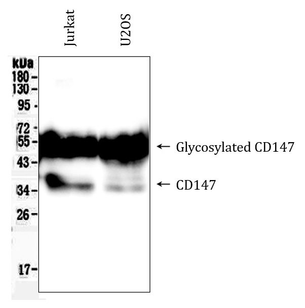
ARG41554 anti-CD147 antibody WB image
Western blot: 50 µg of samples under reducing conditions. Jurkat and U2OS whole cell lysates stained with ARG41554 anti-CD147 antibody at 0.5 µg/ml dilution, overnight at 4°C.
-
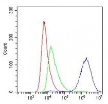
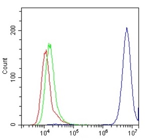
ARG41554 anti-CD147 antibody FACS image
Flow Cytometry: A549 cells were blocked with 10% normal Goat serum and then stained with ARG41554 anti-CD147 antibody (blue) at 1 µg/10^6 cells for 30 min at 20°C, followed by incubation with DyLight®488 labelled secondary antibody. Isotype control antibody (green) was Rabbit IgG (1 µg/10^6 cells) used under the same conditions. Unlabelled sample (red) was also used as a control.
-
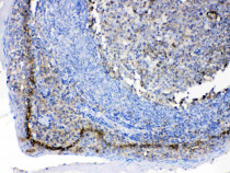
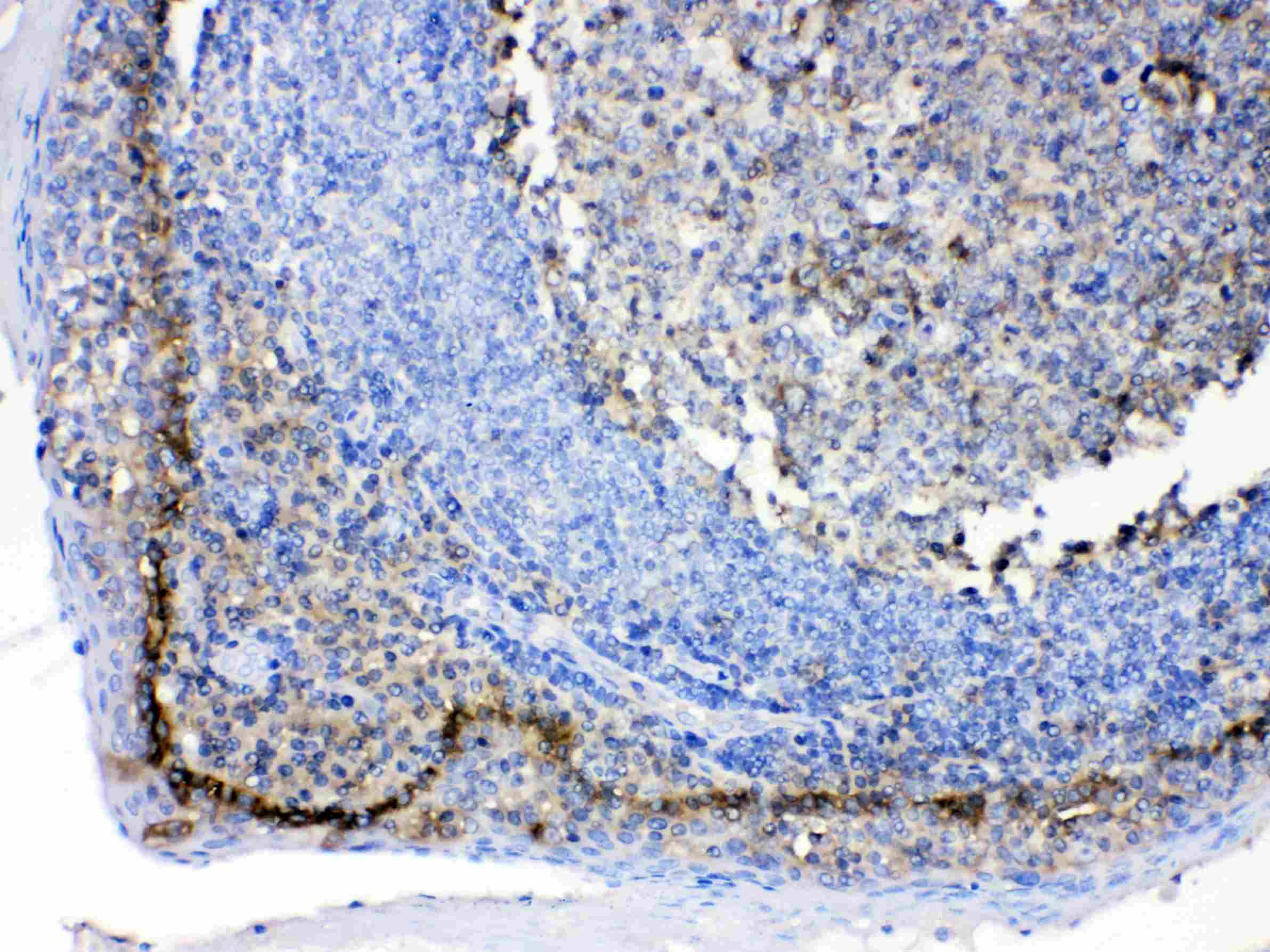
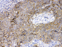
ARG41554 anti-CD147 antibody IHC-P image
Immunohistochemistry: Paraffin-embedded Human tonsil tissue. Antigen Retrieval: Heat mediation was performed in Citrate buffer (pH 6.0, epitope retrieval solution) for 20 min. The tissue section was blocked with 10% goat serum. The tissue section was then stained with ARG41554 anti-CD147 antibody at 1 µg/ml dilution, overnight at 4°C.
-

ARG41554 anti-CD147 antibody IHC-P image
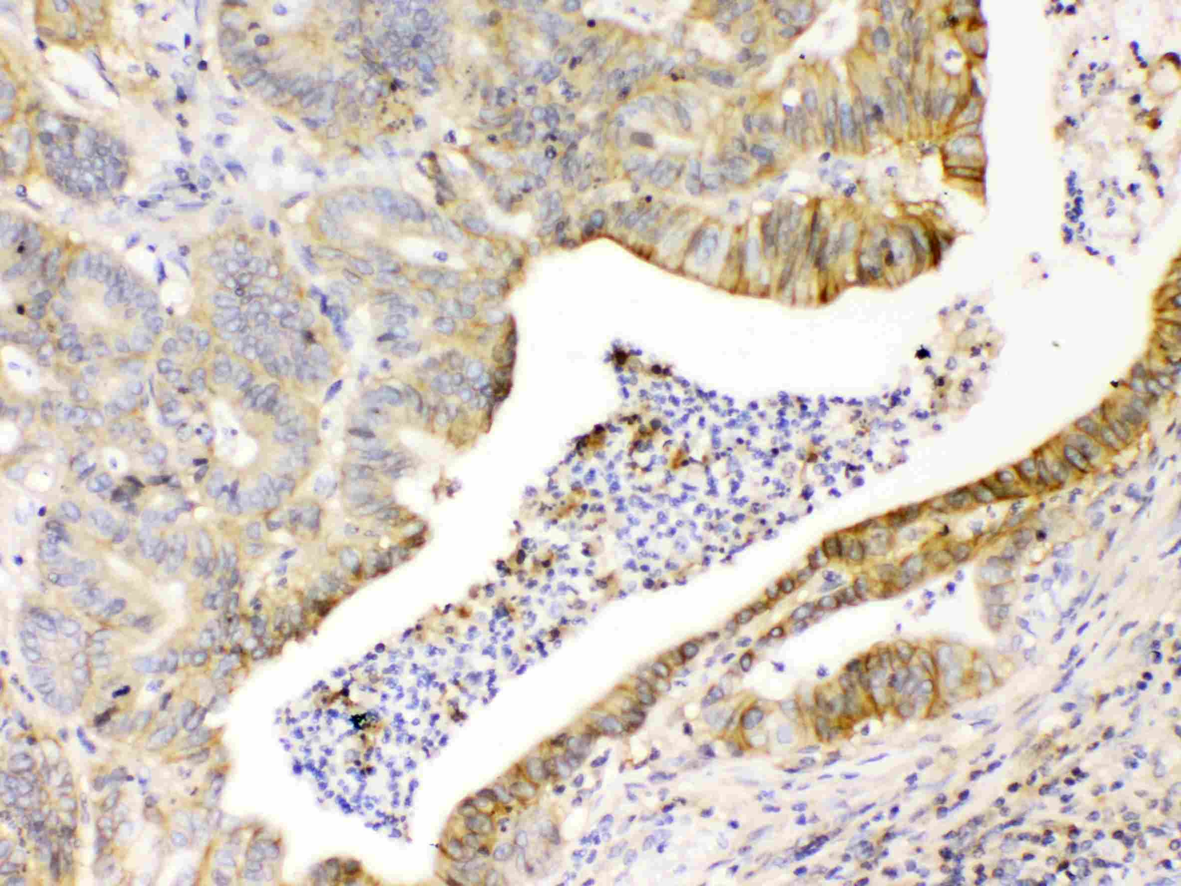
Immunohistochemistry: Paraffin-embedded Human intetsinal cancer tissue. Antigen Retrieval: Heat mediation was performed in Citrate buffer (pH 6.0, epitope retrieval solution) for 20 min. The tissue section was blocked with 10% goat serum. The tissue section was then stained with ARG41554 anti-CD147 antibody at 1 µg/ml dilution, overnight at 4°C.
-
ARG41554 anti-CD147 antibody IHC-P image
Immunohistochemistry: Paraffin-embedded Human intestinal cancer tissue. Antigen Retrieval: Heat mediation was performed in Citrate buffer (pH 6.0, epitope retrieval solution) for 20 min. The tissue section was blocked with 10% goat serum. The tissue section was then stained with ARG41554 anti-CD147 antibody at 1 µg/ml dilution, overnight at 4°C.
-
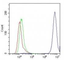
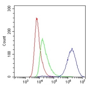
ARG41554 anti-CD147 antibody FACS image
Flow Cytometry: HeLa cells were blocked with 10% normal Goat serum and then stained with ARG41554 anti-CD147 antibody (blue) at 1 µg/10^6 cells for 30 min at 20°C, followed by incubation with DyLight®488 labelled secondary antibody. Isotype control antibody (green) was Rabbit IgG (1 µg/10^6 cells) used under the same conditions. Unlabelled sample (red) was also used as a control.