ARG54192
anti-CD140b / PDGFRB antibody [18A2] (PE)
anti-CD140b / PDGFRB antibody [18A2] (PE) for Flow cytometry and Human
Cancer antibody; Cell Biology and Cellular Response antibody; Signaling Transduction antibody
Overview
| Product Description | PE-conjugated Mouse Monoclonal antibody [18A2] recognizes CD140b / PDGF-RB |
|---|---|
| Tested Reactivity | Hu |
| Tested Application | FACS |
| Specificity | The monoclonal antibody 18A2 recognizes CD140b / PDGFRB, the 180190 kDa beta chain of plateletderived growth factor receptor, which is widely expressed on a variety of mesenchymalderived cells and plays proproliferative roles. |
| Host | Mouse |
| Clonality | Monoclonal |
| Clone | 18A2 |
| Isotype | IgG1 |
| Target Name | CD140b / PDGFRB |
| Immunogen | CD140b-transfected NIH/3T3 cells |
| Conjugation | PE |
| Alternate Names | PDGF-R-beta; IBGC4; CD antigen CD140b; Platelet-derived growth factor receptor beta; CD140B; PDGFR; PDGFR-1; Beta platelet-derived growth factor receptor; PDGFR1; Platelet-derived growth factor receptor 1; PDGFR-beta; CD140 antigen-like family member B; IMF1; EC 2.7.10.1; JTK12; Beta-type platelet-derived growth factor receptor |
Application Instructions
| Application Suggestion |
|
||||
|---|---|---|---|---|---|
| Application Note | * The dilutions indicate recommended starting dilutions and the optimal dilutions or concentrations should be determined by the scientist. |
Properties
| Form | Liquid |
|---|---|
| Purification Note | The purified antibody is conjugated with R-Phycoerythrin (PE) under optimum conditions. The conjugate is purified by size-exclusion chromatography and adjusted for direct use. No reconstitution is necessary. |
| Buffer | PBS, 15 mM Sodium azide and 0.2% (w/v) high-grade protease free BSA |
| Preservative | 15 mM Sodium azide |
| Stabilizer | 0.2% (w/v) high-grade protease free BSA |
| Storage Instruction | Aliquot and store in the dark at 2-8°C. Keep protected from prolonged exposure to light. Avoid repeated freeze/thaw cycles. Suggest spin the vial prior to opening. The antibody solution should be gently mixed before use. |
| Note | For laboratory research only, not for drug, diagnostic or other use. |
Bioinformation
| Database Links |
Swiss-port # P09619 Human Platelet-derived growth factor receptor beta |
|---|---|
| Gene Symbol | PDGFRB |
| Gene Full Name | platelet-derived growth factor receptor, beta polypeptide |
| Background | This gene encodes a cell surface tyrosine kinase receptor for members of the platelet-derived growth factor family. These growth factors are mitogens for cells of mesenchymal origin. The identity of the growth factor bound to a receptor monomer determines whether the functional receptor is a homodimer or a heterodimer, composed of both platelet-derived growth factor receptor alpha and beta polypeptides. This gene is flanked on chromosome 5 by the genes for granulocyte-macrophage colony-stimulating factor and macrophage-colony stimulating factor receptor; all three genes may be implicated in the 5-q syndrome. A translocation between chromosomes 5 and 12, that fuses this gene to that of the translocation, ETV6, leukemia gene, results in chronic myeloproliferative disorder with eosinophilia. [provided by RefSeq, Jul 2008] |
| Function | Tyrosine-protein kinase that acts as cell-surface receptor for homodimeric PDGFB and PDGFD and for heterodimers formed by PDGFA and PDGFB, and plays an essential role in the regulation of embryonic development, cell proliferation, survival, differentiation, chemotaxis and migration. Plays an essential role in blood vessel development by promoting proliferation, migration and recruitment of pericytes and smooth muscle cells to endothelial cells. Plays a role in the migration of vascular smooth muscle cells and the formation of neointima at vascular injury sites. Required for normal development of the cardiovascular system. Required for normal recruitment of pericytes (mesangial cells) in the kidney glomerulus, and for normal formation of a branched network of capillaries in kidney glomeruli. Promotes rearrangement of the actin cytoskeleton and the formation of membrane ruffles. Binding of its cognate ligands - homodimeric PDGFB, heterodimers formed by PDGFA and PDGFB or homodimeric PDGFD -leads to the activation of several signaling cascades; the response depends on the nature of the bound ligand and is modulated by the formation of heterodimers between PDGFRA and PDGFRB. Phosphorylates PLCG1, PIK3R1, PTPN11, RASA1/GAP, CBL, SHC1 and NCK1. Activation of PLCG1 leads to the production of the cellular signaling molecules diacylglycerol and inositol 1,4,5-trisphosphate, mobilization of cytosolic Ca(2+) and the activation of protein kinase C. Phosphorylation of PIK3R1, the regulatory subunit of phosphatidylinositol 3-kinase, leads to the activation of the AKT1 signaling pathway. Phosphorylation of SHC1, or of the C-terminus of PTPN11, creates a binding site for GRB2, resulting in the activation of HRAS, RAF1 and down-stream MAP kinases, including MAPK1/ERK2 and/or MAPK3/ERK1. Promotes phosphorylation and activation of SRC family kinases. Promotes phosphorylation of PDCD6IP/ALIX and STAM. Receptor signaling is down-regulated by protein phosphatases that dephosphorylate the receptor and its down-stream effectors, and by rapid internalization of the activated receptor. [UniProt] |
| Cellular Localization | Cell membrane; Single-pass type I membrane protein. Cytoplasmic vesicle. Lysosome lumen. Note=After ligand binding, the autophosphorylated receptor is ubiquitinated and internalized, leading to its degradation. [UniProt] |
| Research Area | Cancer antibody; Cell Biology and Cellular Response antibody; Signaling Transduction antibody |
| Calculated MW | 124 kDa |
| PTM | Autophosphorylated on tyrosine residues upon ligand binding. Autophosphorylation occurs in trans, i.e. one subunit of the dimeric receptor phosphorylates tyrosine residues on the other subunit. Phosphorylation at Tyr-579, and to a lesser degree, at Tyr-581, is important for interaction with SRC family kinases. Phosphorylation at Tyr-740 and Tyr-751 is important for interaction with PIK3R1. Phosphorylation at Tyr-751 is important for interaction with NCK1. Phosphorylation at Tyr-771 and Tyr-857 is important for interaction with RASA1/GAP. Phosphorylation at Tyr-857 is important for efficient phosphorylation of PLCG1 and PTPN11, resulting in increased phosphorylation of AKT1, MAPK1/ERK2 and/or MAPK3/ERK1, PDCD6IP/ALIX and STAM, and in increased cell proliferation. Phosphorylation at Tyr-1009 is important for interaction with PTPN11. Phosphorylation at Tyr-1009 and Tyr-1021 is important for interaction with PLCG1. Phosphorylation at Tyr-1021 is important for interaction with CBL; PLCG1 and CBL compete for the same binding site. Dephosphorylated by PTPRJ at Tyr-751, Tyr-857, Tyr-1009 and Tyr-1021. Dephosphorylated by PTPN2 at Tyr-579 and Tyr-1021. N-glycosylated. Ubiquitinated. After autophosphorylation, the receptor is polyubiquitinated, leading to its degradation. [UniProt] |
Images (1) Click the Picture to Zoom In
-
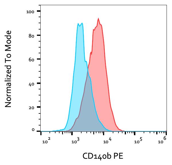
ARG54192 anti-CD140b / PDGFRB antibody [18A2] (PE) FACS image
Flow Cytometry: Human CD140b / PDGFRB stable transfectants (red) and nontransfected cells (blue) stained with ARG54192 anti-CD140b / PDGFRB antibody [18A2] (PE).