ARG44238
anti-CD140a / PDGFRA antibody
anti-CD140a / PDGFRA antibody for Flow cytometry,IHC-Formalin-fixed paraffin-embedded sections,Western blot and Human,Mouse
Overview
| Product Description | Rabbit Polyclonal antibody recognizes CD140a / PDGFRA |
|---|---|
| Tested Reactivity | Hu, Ms |
| Predict Reactivity | Chk, Rat |
| Tested Application | FACS, IHC-P, WB |
| Host | Rabbit |
| Clonality | Polyclonal |
| Isotype | IgG |
| Target Name | CD140a / PDGFRA |
| Antigen Species | Human |
| Immunogen | This CD140a / PDGFRA antibody is generated from rabbits immunized with a KLH conjugated synthetic peptide between 1048-1077 amino acids from the C-terminal region of human CD140a / PDGFRA. |
| Conjugation | Un-conjugated |
Application Instructions
| Application Suggestion |
|
||||||||
|---|---|---|---|---|---|---|---|---|---|
| Application Note | * The dilutions indicate recommended starting dilutions and the optimal dilutions or concentrations should be determined by the scientist. |
Properties
| Form | Liquid |
|---|---|
| Purification | Saturated Ammonium sulphate precipitated |
| Buffer | PBS and 0.09% Sodium azide |
| Preservative | 0.09% Sodium azide |
| Storage Instruction | For continuous use, store undiluted antibody at 2-8°C for up to a week. For long-term storage, aliquot and store at -20°C or below. Storage in frost free freezers is not recommended. Avoid repeated freeze/thaw cycles. Suggest spin the vial prior to opening. The antibody solution should be gently mixed before use. |
| Note | For laboratory research only, not for drug, diagnostic or other use. |
Bioinformation
| Database Links |
Swiss-port # P16234 Human Platelet-derived growth factor receptor alpha Swiss-port # P26618 Mouse Platelet-derived growth factor receptor alpha |
|---|---|
| Gene Symbol | PDGFRA |
| Gene Full Name | platelet-derived growth factor receptor, alpha polypeptide |
| Background | This gene encodes a cell surface tyrosine kinase receptor for members of the platelet-derived growth factor family. These growth factors are mitogens for cells of mesenchymal origin. The identity of the growth factor bound to a receptor monomer determines whether the functional receptor is a homodimer or a heterodimer, composed of both platelet-derived growth factor receptor alpha and beta polypeptides. Studies suggest that this gene plays a role in organ development, wound healing, and tumor progression. Mutations in this gene have been associated with idiopathic hypereosinophilic syndrome, somatic and familial gastrointestinal stromal tumors, and a variety of other cancers. [provided by RefSeq, Mar 2012] |
| Function | Tyrosine-protein kinase that acts as a cell-surface receptor for PDGFA, PDGFB and PDGFC and plays an essential role in the regulation of embryonic development, cell proliferation, survival and chemotaxis. Depending on the context, promotes or inhibits cell proliferation and cell migration. Plays an important role in the differentiation of bone marrow-derived mesenchymal stem cells. Required for normal skeleton development and cephalic closure during embryonic development. Required for normal development of the mucosa lining the gastrointestinal tract, and for recruitment of mesenchymal cells and normal development of intestinal villi. Plays a role in cell migration and chemotaxis in wound healing. Plays a role in platelet activation, secretion of agonists from platelet granules, and in thrombin-induced platelet aggregation. Binding of its cognate ligands - homodimeric PDGFA, homodimeric PDGFB, heterodimers formed by PDGFA and PDGFB or homodimeric PDGFC -leads to the activation of several signaling cascades; the response depends on the nature of the bound ligand and is modulated by the formation of heterodimers between PDGFRA and PDGFRB. Phosphorylates PIK3R1, PLCG1, and PTPN11. Activation of PLCG1 leads to the production of the cellular signaling molecules diacylglycerol and inositol 1,4,5-trisphosphate, mobilization of cytosolic Ca(2+) and the activation of protein kinase C. Phosphorylates PIK3R1, the regulatory subunit of phosphatidylinositol 3-kinase, and thereby mediates activation of the AKT1 signaling pathway. Mediates activation of HRAS and of the MAP kinases MAPK1/ERK2 and/or MAPK3/ERK1. Promotes activation of STAT family members STAT1, STAT3 and STAT5A and/or STAT5B. Receptor signaling is down-regulated by protein phosphatases that dephosphorylate the receptor and its down-stream effectors, and by rapid internalization of the activated receptor. [UniProt] |
| Cellular Localization | Cell membrane; Single-pass type I membrane protein. Note=The activated receptor is rapidly internalized and degraded. [UniProt] |
| Calculated MW | 123 kDa |
| PTM | N-glycosylated. Ubiquitinated, leading to its degradation. Autophosphorylated on tyrosine residues upon ligand binding. Autophosphorylation occurs in trans, i.e. one subunit of the dimeric receptor phosphorylates tyrosine residues on the other subunit. Phosphorylation at Tyr-731 and Tyr-742 is important for interaction with PIK3R1. Phosphorylation at Tyr-720 and Tyr-754 is important for interaction with PTPN11. Phosphorylation at Tyr-762 is important for interaction with CRK. Phosphorylation at Tyr-572 and Tyr-574 is important for interaction with SRC and SRC family members. Phosphorylation at Tyr-988 and Tyr-1018 is important for interaction with PLCG1. [UniProt] |
Images (4) Click the Picture to Zoom In
-
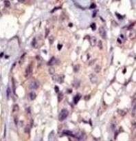
ARG44238 anti-CD140a / PDGFRA antibody IHC-P image
Immunohistochemistry: Human breast carcinoma cancer stained with ARG44238 anti-CD140a / PDGFRA antibody.
-
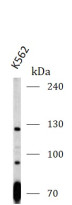
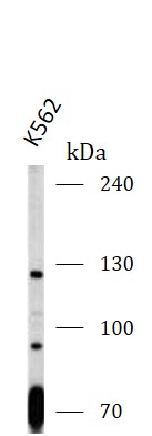
ARG44238 anti-CD140a / PDGFRA antibody WB image
Western blot: K562 stained with ARG44238 anti-CD140a / PDGFRA antibody at 1:1000 dilution.
-
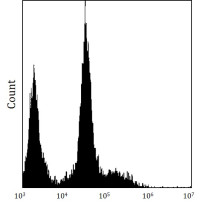
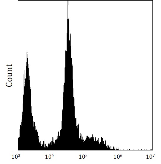
ARG44238 anti-CD140a / PDGFRA antibody FACS image
Flow Cytometry: U251 stained with ARG44238 anti-CD140a / PDGFRA antibody.
-
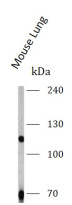
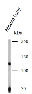
ARG44238 anti-CD140a / PDGFRA antibody WB image
Western blot: Mouse Lung stained with ARG44238 anti-CD140a / PDGFRA antibody at 1:1000 dilution.