ARG65374
anti-CD140a / PDGFRA antibody [16A1] (biotin)
anti-CD140a / PDGFRA antibody [16A1] (biotin) for Flow cytometry and Human
Cancer antibody; Cell Biology and Cellular Response antibody; Signaling Transduction antibody
Overview
| Product Description | Biotin-conjugated Mouse Monoclonal antibody [16A1] recognizes CD140a / PDGF-RA |
|---|---|
| Tested Reactivity | Hu |
| Tested Application | FACS |
| Specificity | The mouse monoclonal antibody 16A1 recognizes CD140a / PDGFRA, the 170 kDa alpha chain of plateletderived growth factor receptor, which is widely expressed on a variety of mesenchymalderived cells and plays proproliferative or antiproliferative roles in various tumours. HLDA VI.; WS Code E022 |
| Host | Mouse |
| Clonality | Monoclonal |
| Clone | 16A1 |
| Isotype | IgG1 |
| Target Name | CD140a / PDGFRA |
| Immunogen | CD140a-transfected NIH 3T3 cells _x000D_ |
| Conjugation | Biotin |
| Alternate Names | Platelet-derived growth factor receptor alpha; PDGFR2; Alpha-type platelet-derived growth factor receptor; RHEPDGFRA; PDGFR-2; Platelet-derived growth factor receptor 2; CD140A; Platelet-derived growth factor alpha receptor; CD140 antigen-like family member A; CD antigen CD140a; PDGF-R-alpha; Alpha platelet-derived growth factor receptor; CD140a antigen; EC 2.7.10.1; PDGFR-alpha |
Application Instructions
| Application Suggestion |
|
||||
|---|---|---|---|---|---|
| Application Note | * The dilutions indicate recommended starting dilutions and the optimal dilutions or concentrations should be determined by the scientist. |
Properties
| Form | Liquid |
|---|---|
| Purification Note | The purified antibody is conjugated with Biotin-LC-NHS under optimum conditions. The reagent is free of unconjugated biotin. |
| Buffer | PBS (pH 7.4) and 15 mM Sodium azide |
| Preservative | 15 mM Sodium azide |
| Concentration | 1 mg/ml |
| Storage Instruction | Aliquot and store in the dark at 2-8°C. Keep protected from prolonged exposure to light. Avoid repeated freeze/thaw cycles. Suggest spin the vial prior to opening. The antibody solution should be gently mixed before use. |
| Note | For laboratory research only, not for drug, diagnostic or other use. |
Bioinformation
| Database Links |
Swiss-port # P16234 Human Platelet-derived growth factor receptor alpha |
|---|---|
| Gene Symbol | PDGFRA |
| Gene Full Name | platelet-derived growth factor receptor, alpha polypeptide |
| Background | CD140a / PDGF-RA (platelet-derived growth factor receptor alpha) is a cell surface receptor for members of platelet-derived growth factor family, whose intracellular part contains a tyrosine kinase domain. CD140a forms homodimers, or heterodimerizes with CD140b / PDGF-RB. Whereas CD140b induces in different cell types their proliferation and migration, the role of CD140a is more controversial, with pro-proliferative or anti-proliferative effects. CD140a has early developmental functions, mediates mesodermal cell migration, and later acts in signaling associated in epithelial-mesenchymal interactions. |
| Function | Tyrosine-protein kinase that acts as a cell-surface receptor for PDGFA, PDGFB and PDGFC and plays an essential role in the regulation of embryonic development, cell proliferation, survival and chemotaxis. Depending on the context, promotes or inhibits cell proliferation and cell migration. Plays an important role in the differentiation of bone marrow-derived mesenchymal stem cells. Required for normal skeleton development and cephalic closure during embryonic development. Required for normal development of the mucosa lining the gastrointestinal tract, and for recruitment of mesenchymal cells and normal development of intestinal villi. Plays a role in cell migration and chemotaxis in wound healing. Plays a role in platelet activation, secretion of agonists from platelet granules, and in thrombin-induced platelet aggregation. Binding of its cognate ligands - homodimeric PDGFA, homodimeric PDGFB, heterodimers formed by PDGFA and PDGFB or homodimeric PDGFC -leads to the activation of several signaling cascades; the response depends on the nature of the bound ligand and is modulated by the formation of heterodimers between PDGFRA and PDGFRB. Phosphorylates PIK3R1, PLCG1, and PTPN11. Activation of PLCG1 leads to the production of the cellular signaling molecules diacylglycerol and inositol 1,4,5-trisphosphate, mobilization of cytosolic Ca(2+) and the activation of protein kinase C. Phosphorylates PIK3R1, the regulatory subunit of phosphatidylinositol 3-kinase, and thereby mediates activation of the AKT1 signaling pathway. Mediates activation of HRAS and of the MAP kinases MAPK1/ERK2 and/or MAPK3/ERK1. Promotes activation of STAT family members STAT1, STAT3 and STAT5A and/or STAT5B. Receptor signaling is down-regulated by protein phosphatases that dephosphorylate the receptor and its down-stream effectors, and by rapid internalization of the activated receptor. [UniProt] |
| Research Area | Cancer antibody; Cell Biology and Cellular Response antibody; Signaling Transduction antibody |
| Calculated MW | 123 kDa |
| PTM | N-glycosylated. Ubiquitinated, leading to its degradation. Autophosphorylated on tyrosine residues upon ligand binding. Autophosphorylation occurs in trans, i.e. one subunit of the dimeric receptor phosphorylates tyrosine residues on the other subunit. Phosphorylation at Tyr-731 and Tyr-742 is important for interaction with PIK3R1. Phosphorylation at Tyr-720 and Tyr-754 is important for interaction with PTPN11. Phosphorylation at Tyr-762 is important for interaction with CRK. Phosphorylation at Tyr-572 and Tyr-574 is important for interaction with SRC and SRC family members. Phosphorylation at Tyr-988 and Tyr-1018 is important for interaction with PLCG1. |
Images (1) Click the Picture to Zoom In
-
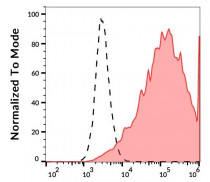
ARG65374 anti-CD140a / PDGFRA antibody [16A1] (biotin) FACS image
Flow Cytometry: CD140a-transfected cells stained with ARG65374 anti-CD140a / PDGFRA antibody [16A1] (biotin) (red-filled) at 2 µg/ml dilution, followed by Streptavidin (APC). Blank sample (black-dashed).


