ARG42300
anti-CD135 / FLT3 antibody [BV10A4] (APC)
anti-CD135 / FLT3 antibody [BV10A4] (APC) for Flow cytometry and Human
Overview
| Product Description | APC-conjugated Mouse Monoclonal antibody [BV10A4] recognizes CD135 / FLT3 |
|---|---|
| Tested Reactivity | Hu |
| Species Does Not React With | Ms |
| Tested Application | FACS |
| Specificity | The mouse monoclonal antibody BV10A4 (BV10) reacts with an extracellular epitope of CD135 (FLT3, FLK2, STK-1), a 130-160 kDa type I transmembrane receptor tyrosine kinase that is involved in early steps of hematopoiesis. |
| Host | Mouse |
| Clonality | Monoclonal |
| Clone | BV10A4 |
| Isotype | IgG1 |
| Target Name | CD135 / FLT3 |
| Antigen Species | Human |
| Immunogen | BV-173 leukemic cell line. |
| Conjugation | APC |
| Alternate Names | CD135; FLK2; Receptor-type tyrosine-protein kinase FLT3; FLK-2; STK-1; STK1; FL cytokine receptor; FLT-3; Stem cell tyrosine kinase 1; Fetal liver kinase-2; Fms-like tyrosine kinase 3; CD antigen CD135; EC 2.7.10.1 |
Application Instructions
| Application Suggestion |
|
||||
|---|---|---|---|---|---|
| Application Note | * The dilutions indicate recommended starting dilutions and the optimal dilutions or concentrations should be determined by the scientist. |
Properties
| Form | Liquid |
|---|---|
| Purification | Purified |
| Buffer | PBS and 15 mM Sodium azide. |
| Preservative | 15 mM Sodium azide |
| Storage Instruction | Aliquot and store in the dark at 2-8°C. Keep protected from prolonged exposure to light. Avoid repeated freeze/thaw cycles. Suggest spin the vial prior to opening. The antibody solution should be gently mixed before use. |
| Note | For laboratory research only, not for drug, diagnostic or other use. |
Bioinformation
| Database Links |
Swiss-port # P36888 Human Receptor-type tyrosine-protein kinase FLT3 |
|---|---|
| Gene Symbol | FLT3 |
| Gene Full Name | fms-related tyrosine kinase 3 |
| Background | This gene encodes a class III receptor tyrosine kinase that regulates hematopoiesis. This receptor is activated by binding of the fms-related tyrosine kinase 3 ligand to the extracellular domain, which induces homodimer formation in the plasma membrane leading to autophosphorylation of the receptor. The activated receptor kinase subsequently phosphorylates and activates multiple cytoplasmic effector molecules in pathways involved in apoptosis, proliferation, and differentiation of hematopoietic cells in bone marrow. Mutations that result in the constitutive activation of this receptor result in acute myeloid leukemia and acute lymphoblastic leukemia. [provided by RefSeq, Jan 2015] |
| Function | Tyrosine-protein kinase that acts as cell-surface receptor for the cytokine FLT3LG and regulates differentiation, proliferation and survival of hematopoietic progenitor cells and of dendritic cells. Promotes phosphorylation of SHC1 and AKT1, and activation of the downstream effector MTOR. Promotes activation of RAS signaling and phosphorylation of downstream kinases, including MAPK1/ERK2 and/or MAPK3/ERK1. Promotes phosphorylation of FES, FER, PTPN6/SHP, PTPN11/SHP-2, PLCG1, and STAT5A and/or STAT5B. Activation of wild-type FLT3 causes only marginal activation of STAT5A or STAT5B. Mutations that cause constitutive kinase activity promote cell proliferation and resistance to apoptosis via the activation of multiple signaling pathways. [UniProt] |
| Cellular Localization | Membrane; Single-pass type I membrane protein. Endoplasmic reticulum lumen. Note=Constitutively activated mutant forms with internal tandem duplications are less efficiently transported to the cell surface and a significant proportion is retained in an immature form in the endoplasmic reticulum lumen. The activated kinase is rapidly targeted for degradation. [UniProt] |
| Calculated MW | 113 kDa |
| PTM | N-glycosylated, contains complex N-glycans with sialic acid. Autophosphorylated on several tyrosine residues in response to FLT3LG binding. FLT3LG binding also increases phosphorylation of mutant kinases that are constitutively activated. Dephosphorylated by PTPRJ/DEP-1, PTPN1, PTPN6/SHP-1, and to a lesser degree by PTPN12. Dephosphorylation is important for export from the endoplasmic reticulum and location at the cell membrane. Rapidly ubiquitinated by UBE2L6 and the E3 ubiquitin-protein ligase SIAH1 after autophosphorylation, leading to its proteasomal degradation. [UniProt] |
Images (2) Click the Picture to Zoom In
-

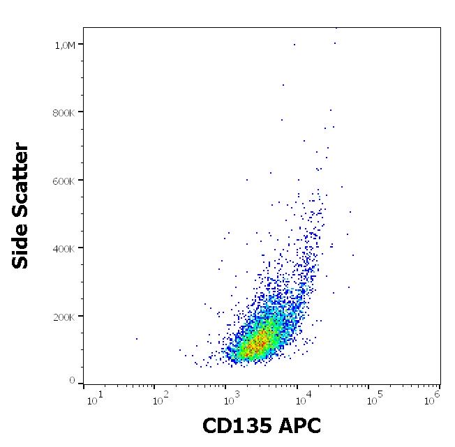
ARG42300 anti-CD135 / FLT3 antibody [BV10A4] (APC) FACS image
Flow Cytometry: REH cell suspension stained with ARG42300 anti-CD135 / FLT3 antibody [BV10A4] (APC) at 10 µl / 10^6 cells in 100 µl of cell suspension.
-

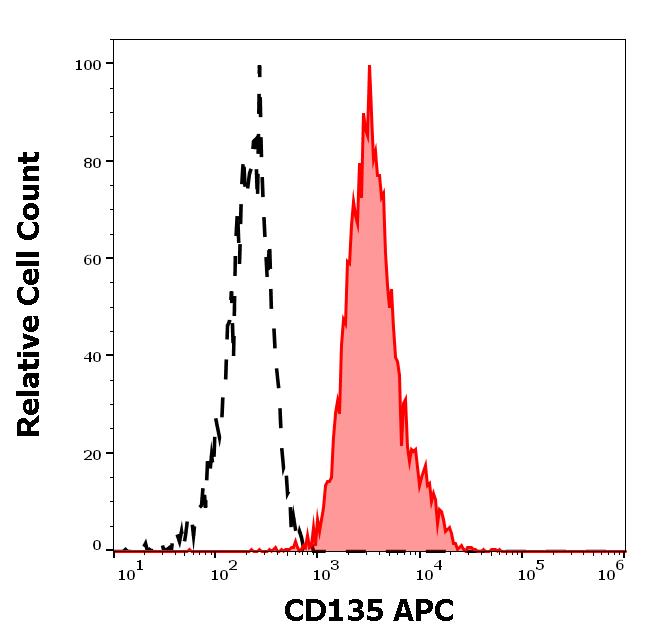
ARG42300 anti-CD135 / FLT3 antibody [BV10A4] (APC) FACS image
Flow Cytometry: Separation of REH cells stained with ARG42300 anti-CD135 / FLT3 antibody [BV10A4] (APC) at 10 µl / 10^6 cells in 100 µl of cell suspension (red-filled) from REH cells stained with ARG65336 Mouse IgG1 Kappa Isotype Control antibody [MOPC-21] (APC) at 5 µg/ml dilution (black-dashed).