ARG51168
anti-CD135 / FLT3 antibody
anti-CD135 / FLT3 antibody for ICC/IF,Western blot and Human
Immune System antibody; Signaling Transduction antibody

1
Overview
| Product Description | Rabbit Polyclonal antibody recognizes CD135 / FLT3 |
|---|---|
| Tested Reactivity | Hu |
| Tested Application | ICC/IF, WB |
| Host | Rabbit |
| Clonality | Polyclonal |
| Isotype | IgG |
| Target Name | CD135 / FLT3 |
| Antigen Species | Human |
| Immunogen | Peptide sequence around aa.589~593 (Y-F-Y-V-D) derived from Human CD135 / Flt3. |
| Conjugation | Un-conjugated |
| Alternate Names | CD135; FLK2; Receptor-type tyrosine-protein kinase FLT3; FLK-2; STK-1; STK1; FL cytokine receptor; FLT-3; Stem cell tyrosine kinase 1; Fetal liver kinase-2; Fms-like tyrosine kinase 3; CD antigen CD135; EC 2.7.10.1 |
Application Instructions
| Application Suggestion |
|
||||||
|---|---|---|---|---|---|---|---|
| Application Note | * The dilutions indicate recommended starting dilutions and the optimal dilutions or concentrations should be determined by the scientist. |
Properties
| Form | Liquid |
|---|---|
| Purification | Antibodies were produced by immunizing rabbits with KLH-conjugated synthetic peptide. Antibodies were purified by affinity-chromatography using epitope-specific peptide. |
| Buffer | PBS (without Mg2+ and Ca2+, pH 7.4), 150mM NaCl, 0.02% Sodium azide and 50% Glycerol. |
| Preservative | 0.02% Sodium azide |
| Stabilizer | 50% Glycerol |
| Concentration | 1 mg/ml |
| Storage Instruction | For continuous use, store undiluted antibody at 2-8°C for up to a week. For long-term storage, aliquot and store at -20°C. Storage in frost free freezers is not recommended. Avoid repeated freeze/thaw cycles. Suggest spin the vial prior to opening. The antibody solution should be gently mixed before use. |
| Note | For laboratory research only, not for drug, diagnostic or other use. |
Bioinformation
| Database Links |
Swiss-port # P36888 Human Receptor-type tyrosine-protein kinase FLT3 |
|---|---|
| Gene Symbol | FLT3 |
| Gene Full Name | fms-related tyrosine kinase 3 |
| Background | FLT3 encodes a class III receptor tyrosine kinase that regulates hematopoiesis. The receptor consists of an extracellular domain composed of five immunoglobulin-like domains, one transmembrane region, and a cytoplasmic kinase domain split into two parts by a kinase-insert domain. The receptor is activated by binding of the fms-related tyrosine kinase 3 ligand to the extracellular domain, which induces homodimer formation in the plasma membrane leading to autophosphorylation of the receptor. The activated receptor kinase subsequently phosphorylates and activates multiple cytoplasmic effector molecules in pathways involved in apoptosis, proliferation, and differentiation of hematopoietic cells in bone marrow. Mutations that result in the constitutive activation of this receptor result in acute myeloid leukemia and acute lymphoblastic leukemia. |
| Function | Tyrosine-protein kinase that acts as cell-surface receptor for the cytokine FLT3LG and regulates differentiation, proliferation and survival of hematopoietic progenitor cells and of dendritic cells. Promotes phosphorylation of SHC1 and AKT1, and activation of the downstream effector MTOR. Promotes activation of RAS signaling and phosphorylation of downstream kinases, including MAPK1/ERK2 and/or MAPK3/ERK1. Promotes phosphorylation of FES, FER, PTPN6/SHP, PTPN11/SHP-2, PLCG1, and STAT5A and/or STAT5B. Activation of wild-type FLT3 causes only marginal activation of STAT5A or STAT5B. Mutations that cause constitutive kinase activity promote cell proliferation and resistance to apoptosis via the activation of multiple signaling pathways. [UniProt] |
| Research Area | Immune System antibody; Signaling Transduction antibody |
| Calculated MW | 113 kDa |
| PTM | N-glycosylated, contains complex N-glycans with sialic acid. Autophosphorylated on several tyrosine residues in response to FLT3LG binding. FLT3LG binding also increases phosphorylation of mutant kinases that are constitutively activated. Dephosphorylated by PTPRJ/DEP-1, PTPN1, PTPN6/SHP-1, and to a lesser degree by PTPN12. Dephosphorylation is important for export from the endoplasmic reticulum and location at the cell membrane. Rapidly ubiquitinated by UBE2L6 and the E3 ubiquitin-protein ligase SIAH1 after autophosphorylation, leading to its proteasomal degradation. |
Images (2) Click the Picture to Zoom In
-

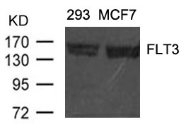
ARG51168 anti-CD135 / FLT3 antibody WB image
Western blot: Extracts from 293 and MCF cells stained with ARG51168 anti-CD135 / FLT3 antibody.
-

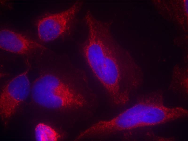
ARG51168 anti-CD135 / FLT3 antibody ICC/IF image
Immunofluorescence: methanol-fixed HeLa cells stained with ARG51168 anti-CD135 / FLT3 antibody.
Customer's Feedback
Good
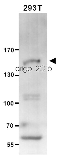
Review for anti-CD135 / FLT3 antibody
Application:WB
Sample:293T
Sample Loading Amount:20 µg
Primary Antibody Dilution Factor:1:500
Primary Antibody Incubation Time:overnight
Primary Antibody Incubation Temperature:4 ºC


