ARG22993
anti-CD13 antibody [R3-63]
anti-CD13 antibody [R3-63] for Flow cytometry,ICC/IF,IHC-Frozen sections,IHC-Formalin-fixed paraffin-embedded sections and Mouse
Overview
| Product Description | Rat Monoclonal antibody [R3-63] recognizes CD13 Rat anti Mouse CD13 antibody, clone R3-63 recognizes mouse aminopeptidase N (APN), a cell surface protein homologous with human CD13. In the mouse, CD13 is a non-covalently linked homodimer of approximately 150 kDa subunits expressed by a variety of cells including monocytes, macrophages, dendritic cell and veiled cells.Rat anti Mouse CD13 antibody, clone R3-63 has been reported to block mouse APN enzyme activity (Hansen et al. 1993). |
|---|---|
| Tested Reactivity | Ms |
| Tested Application | FACS, ICC/IF, IHC-Fr, IHC-P |
| Host | Rat |
| Clonality | Monoclonal |
| Clone | R3-63 |
| Isotype | IgG2a |
| Target Name | CD13 |
| Antigen Species | Mouse |
| Immunogen | Mouse intestinal APN |
| Conjugation | Un-conjugated |
| Alternate Names | AP-N; PEPN; LAP1; CD antigen CD13; Aminopeptidase M; gp150; Aminopeptidase N; EC 3.4.11.2; Myeloid plasma membrane glycoprotein CD13; APN; CD13; P150; AP-M; GP150; hAPN; Microsomal aminopeptidase; Alanyl aminopeptidase |
Application Instructions
| Application Suggestion |
|
||||||||||
|---|---|---|---|---|---|---|---|---|---|---|---|
| Application Note | IHC-P: Antigen Retrieval: Boil tissue section in Sodium citrate buffer (pH 6.0). FACS: Use 10 µl of the suggested working dilution to label 10^6 cells in 100 µl. * The dilutions indicate recommended starting dilutions and the optimal dilutions or concentrations should be determined by the scientist. |
Properties
| Form | Liquid |
|---|---|
| Purification | Purification with Protein G. |
| Buffer | PBS and 0.09% Sodium azide |
| Preservative | 0.09% Sodium azide |
| Concentration | 1 mg/ml |
| Storage Instruction | For continuous use, store undiluted antibody at 2-8°C for up to a week. For long-term storage, aliquot and store at -20°C or below. Storage in frost free freezers is not recommended. Avoid repeated freeze/thaw cycles. Suggest spin the vial prior to opening. The antibody solution should be gently mixed before use. |
| Note | For laboratory research only, not for drug, diagnostic or other use. |
Bioinformation
| Database Links | |
|---|---|
| Gene Symbol | Anpep |
| Gene Full Name | alanyl (membrane) aminopeptidase |
| Background | Aminopeptidase N is located in the small-intestinal and renal microvillar membrane, and also in other plasma membranes. In the small intestine aminopeptidase N plays a role in the final digestion of peptides generated from hydrolysis of proteins by gastric and pancreatic proteases. Its function in proximal tubular epithelial cells and other cell types is less clear. The large extracellular carboxyterminal domain contains a pentapeptide consensus sequence characteristic of members of the zinc-binding metalloproteinase superfamily. Sequence comparisons with known enzymes of this class showed that CD13 and aminopeptidase N are identical. The latter enzyme was thought to be involved in the metabolism of regulatory peptides by diverse cell types, including small intestinal and renal tubular epithelial cells, macrophages, granulocytes, and synaptic membranes from the CNS. Human aminopeptidase N is a receptor for one strain of human coronavirus that is an important cause of upper respiratory tract infections. Defects in this gene appear to be a cause of various types of leukemia or lymphoma. [provided by RefSeq, Jul 2008] |
| Function | Broad specificity aminopeptidase. Plays a role in the final digestion of peptides generated from hydrolysis of proteins by gastric and pancreatic proteases. May play a critical role in the pathogenesis of cholesterol gallstone disease. May be involved in the metabolism of regulatory peptides of diverse cell types, responsible for the processing of peptide hormones, such as angiotensin III and IV, neuropeptides, and chemokines. Found to cleave antigen peptides bound to major histocompatibility complex class II molecules of presenting cells and to degrade neurotransmitters at synaptic junctions. Is also implicated as a regulator of IL-8 bioavailability in the endometrium, and therefore may contribute to the regulation of angiogenesis. Is used as a marker for acute myeloid leukemia and plays a role in tumor invasion. In case of human coronavirus 229E (HCoV-229E) infection, serves as receptor for HCoV-229E spike glycoprotein. Mediates as well human cytomegalovirus (HCMV) infection. [UniProt] |
| Calculated MW | 110 kDa |
| PTM | Sulfated. N- and O-glycosylated. May undergo proteolysis and give rise to a soluble form. |
Images (6) Click the Picture to Zoom In
-
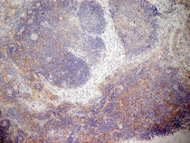
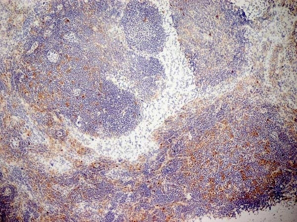
ARG22993 anti-CD13 antibody [R3-63] IHC-Fr image
Immunohistochemistry: Mouse lymph node cryosection stained with ARG22993 anti-CD13 antibody [R3-63] followed by HRP-conjugated Goat anti Rat IgG antibody. (Low power).
-
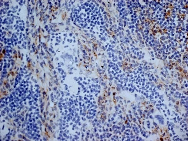
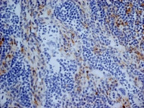
ARG22993 anti-CD13 antibody [R3-63] IHC-Fr image
Immunohistochemistry: Mouse lymph node cryosection stained with ARG22993 anti-CD13 antibody [R3-63] followed by HRP-conjugated Goat anti Rat IgG antibody. (Medium power).
-
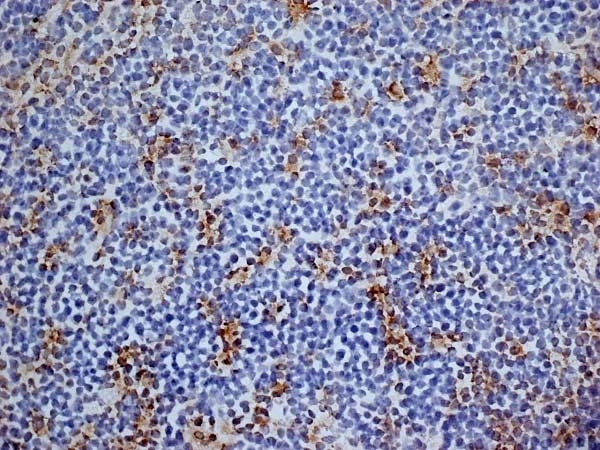
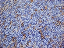
ARG22993 anti-CD13 antibody [R3-63] IHC-Fr image
Immunohistochemistry: Mouse lymph node cryosection stained with ARG22993 anti-CD13 antibody [R3-63] followed by HRP-conjugated Goat anti Rat IgG antibody. (High power).
-
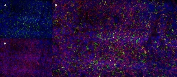
ARG22993 anti-CD13 antibody [R3-63] IHC-Fr image
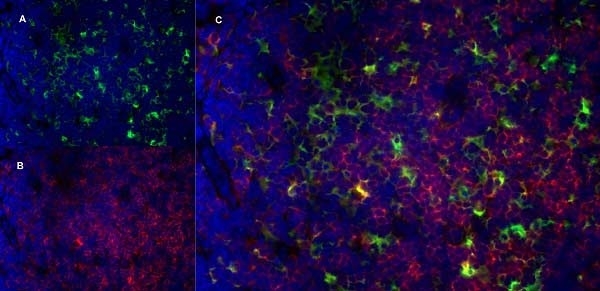
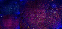
Immunohistochemistry: Mouse lymph node cryosection stained with ARG22993 anti-CD13 antibody [R3-63], green in A and Rat anti Mouse CD8, clone YTS105.18, red in B. C is the merged image with nuclei counterstained blue using DAPI. (Low power).
-
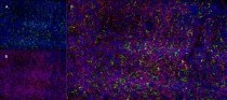
ARG22993 anti-CD13 antibody [R3-63] IHC-Fr image
Immunohistochemistry: Mouse lymph node cryosection stained with ARG22993 anti-CD13 antibody [R3-63], green in A and Rat anti Mouse CD8, clone YTS105.18, red in B. C is the merged image with nuclei counterstained blue using DAPI. (Medium power).
-

ARG22993 anti-CD13 antibody [R3-63] IHC-Fr image
Immunohistochemistry: Mouse lymph node cryosection stained with ARG22993 anti-CD13 antibody [R3-63], green in A and Rat anti Mouse CD8, clone YTS105.18, red in B. C is the merged image with nuclei counterstained blue using DAPI. (High power).