ARG42298
anti-CD120a / TNFR1 antibody [H398] (PE)
anti-CD120a / TNFR1 antibody [H398] (PE) for Flow cytometry and Human
Overview
| Product Description | PE-conjugated Mouse Monoclonal antibody [H398] recognizes CD120a / TNFR1 |
|---|---|
| Tested Reactivity | Hu |
| Tested Application | FACS |
| Specificity | The mouse monoclonal antibody H398 recognizes the extracellular domain of CD120a, a 55 kDa receptor for tumor necrosis factor. The antibody blocks biological activity of both natural and recombinant human TNF alpha and TNF beta. |
| Host | Mouse |
| Clonality | Monoclonal |
| Clone | H398 |
| Isotype | IgG2a |
| Target Name | CD120a / TNFR1 |
| Antigen Species | Human |
| Immunogen | Recombinant full length Human CD120a. |
| Conjugation | PE |
| Alternate Names | TNF-R; p60; TNFAR; CD antigen CD120a; TNFR55; TBP1; TNF-RI; TNFR1-d2; Tumor necrosis factor receptor superfamily member 1A; FPF; TNFR60; CD120a; TNFR1; p55; TNF-R55; TNF-R-I; MS5; TNFR-I; Tumor necrosis factor receptor 1; TBPI; Tumor necrosis factor receptor type I; TNF-R1; p55-R |
Application Instructions
| Application Suggestion |
|
||||
|---|---|---|---|---|---|
| Application Note | * The dilutions indicate recommended starting dilutions and the optimal dilutions or concentrations should be determined by the scientist. |
Properties
| Form | Liquid |
|---|---|
| Purification | Purified |
| Buffer | PBS and 15 mM Sodium azide. |
| Preservative | 15 mM Sodium azide |
| Storage Instruction | Aliquot and store in the dark at 2-8°C. Keep protected from prolonged exposure to light. Avoid repeated freeze/thaw cycles. Suggest spin the vial prior to opening. The antibody solution should be gently mixed before use. |
| Note | For laboratory research only, not for drug, diagnostic or other use. |
Bioinformation
| Database Links |
Swiss-port # P19438 Human Tumor necrosis factor receptor superfamily member 1A |
|---|---|
| Gene Symbol | TNFRSF1A |
| Gene Full Name | tumor necrosis factor receptor superfamily, member 1A |
| Background | This gene encodes a member of the TNF receptor superfamily of proteins. The encoded receptor is found in membrane-bound and soluble forms that interact with membrane-bound and soluble forms, respectively, of its ligand, tumor necrosis factor alpha. Binding of membrane-bound tumor necrosis factor alpha to the membrane-bound receptor induces receptor trimerization and activation, which plays a role in cell survival, apoptosis, and inflammation. Proteolytic processing of the encoded receptor results in release of the soluble form of the receptor, which can interact with free tumor necrosis factor alpha to inhibit inflammation. Mutations in this gene underlie tumor necrosis factor receptor-associated periodic syndrome (TRAPS), characterized by fever, abdominal pain and other features. Mutations in this gene may also be associated with multiple sclerosis in human patients. [provided by RefSeq, Sep 2016] |
| Function | Receptor for TNFSF2/TNF-alpha and homotrimeric TNFSF1/lymphotoxin-alpha. The adapter molecule FADD recruits caspase-8 to the activated receptor. The resulting death-inducing signaling complex (DISC) performs caspase-8 proteolytic activation which initiates the subsequent cascade of caspases (aspartate-specific cysteine proteases) mediating apoptosis. Contributes to the induction of non-cytocidal TNF effects including anti-viral state and activation of the acid sphingomyelinase. [UniProt] |
| Cellular Localization | Cell membrane; Single-pass type I membrane protein. Golgi apparatus membrane; Single-pass type I membrane protein. Secreted. Note=A secreted form is produced through proteolytic processing. Isoform 4: Secreted. Note=Lacks a Golgi-retention motif, is not membrane bound and therefore is secreted. [UniProt] |
| Calculated MW | 50 kDa |
| PTM | The soluble form is produced from the membrane form by proteolytic processing. [UniProt] |
Images (2) Click the Picture to Zoom In
-

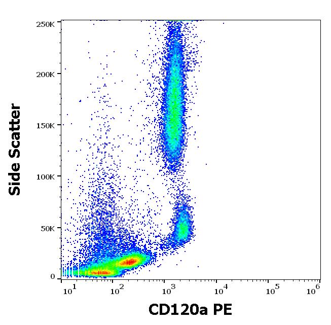
ARG42298 anti-CD120a / TNFR1 antibody [H398] (PE) FACS image
Flow Cytometry: Human peripheral whole blood stained with ARG42298 anti-CD120a / TNFR1 antibody [H398] (PE) at 10 µl / 100 µl of peripheral whole blood.
-
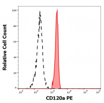
ARG42298 anti-CD120a / TNFR1 antibody [H398] (PE) FACS image
Flow Cytometry: Separation of Human monocytes (red-filled) from lymphocytes (black-dashed). Human peripheral whole blood stained with ARG42298 anti-CD120a / TNFR1 antibody [H398] (PE) at 10 µl / 100 µl of peripheral whole blood.