ARG56617
anti-CCL2 / MCP1 antibody
anti-CCL2 / MCP1 antibody for ELISA,IHC-Formalin-fixed paraffin-embedded sections,Western blot and Human,Mouse
Overview
| Product Description | Rabbit Polyclonal antibody recognizes CCL2 / MCP1 |
|---|---|
| Tested Reactivity | Hu, Ms |
| Tested Application | ELISA, IHC-P, WB |
| Host | Rabbit |
| Clonality | Polyclonal |
| Isotype | IgG |
| Target Name | CCL2 / MCP1 |
| Antigen Species | Human |
| Immunogen | E.coli derived Recombinant Human CCL2 / MCP1. (QPDAINAPVT CCYNFTNRKI SVQRLASYRR ITSSKCPKEA VIFKTIVAKE ICADPKQKWV QDSMDHLDKQ TQTPKT) |
| Conjugation | Un-conjugated |
| Alternate Names | MCP1; Monocyte chemotactic and activating factor; MCAF; Monocyte chemotactic protein 1; Monocyte secretory protein JE; HSMCR30; Small-inducible cytokine A2; HC11; SMC-CF; GDCF-2; SCYA2; C-C motif chemokine 2; Monocyte chemoattractant protein 1; MCP-1 |
Application Instructions
| Application Suggestion |
|
||||||||
|---|---|---|---|---|---|---|---|---|---|
| Application Note | * The dilutions indicate recommended starting dilutions and the optimal dilutions or concentrations should be determined by the scientist. |
Properties
| Form | Liquid |
|---|---|
| Purification | Affinity purification with immunogen. |
| Buffer | PBS (pH 7.2) |
| Concentration | 1 mg/ml |
| Storage Instruction | For continuous use, store undiluted antibody at 2-8°C for up to a week. For long-term storage, aliquot and store at -20°C or below. Storage in frost free freezers is not recommended. Avoid repeated freeze/thaw cycles. Suggest spin the vial prior to opening. The antibody solution should be gently mixed before use. |
| Note | For laboratory research only, not for drug, diagnostic or other use. |
Bioinformation
| Database Links | |
|---|---|
| Gene Symbol | CCL2 |
| Gene Full Name | chemokine (C-C motif) ligand 2 |
| Background | This gene is one of several cytokine genes clustered on the q-arm of chromosome 17. Chemokines are a superfamily of secreted proteins involved in immunoregulatory and inflammatory processes. The superfamily is divided into four subfamilies based on the arrangement of N-terminal cysteine residues of the mature peptide. This chemokine is a member of the CC subfamily which is characterized by two adjacent cysteine residues. This cytokine displays chemotactic activity for monocytes and basophils but not for neutrophils or eosinophils. It has been implicated in the pathogenesis of diseases characterized by monocytic infiltrates, like psoriasis, rheumatoid arthritis and atherosclerosis. It binds to chemokine receptors CCR2 and CCR4. [provided by RefSeq, Jul 2013] |
| Function | Chemotactic factor that attracts monocytes and basophils but not neutrophils or eosinophils. Augments monocyte anti-tumor activity. Has been implicated in the pathogenesis of diseases characterized by monocytic infiltrates, like psoriasis, rheumatoid arthritis or atherosclerosis. May be involved in the recruitment of monocytes into the arterial wall during the disease process of atherosclerosis. [UniProt] |
| Highlight | Related products: MCP1 antibodies; MCP1 ELISA Kits; MCP1 Duos / Panels; Anti-Rabbit IgG secondary antibodies; Related news: HMGB1 in inflammation Inflammatory Cytokines |
| Calculated MW | 11 kDa (Human); 16 kDa (Mouse) |
| PTM | Processing at the N-terminus can regulate receptor and target cell selectivity. Deletion of the N-terminal residue converts it from an activator of basophil to an eosinophil chemoattractant. |
Images (6) Click the Picture to Zoom In
-
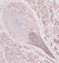
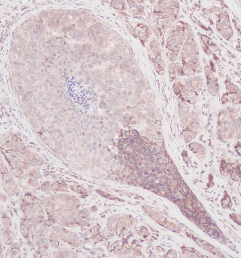
ARG56617 anti-CCL2 / MCP1 antibody IHC-P image
Immunohistochemistry: Formalin-fixed and paraffin-embedded sections of Human breast invasive ductal carcinoma. The recommended ARG56617 anti-CCL2 / MCP1 antibody concentration is 2.5 µg/ml and a two-hour incubation at RT. An HRP-labeled polymer detection system was used with a DAB chromogen. Antigen Retrieval: Boil tissue section in Sodium Citrate buffer (pH 6.0) followed by cooling at RT for 20 min.
-

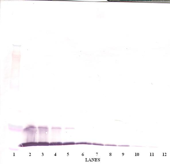
ARG56617 anti-CCL2 / MCP1 antibody WB image
Western blot: 250 - 0.24 ng of Human MCP-1(MCAF) stained with ARG56617 anti-CCL2 / MCP1 antibody, under reducing conditions.
-
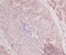
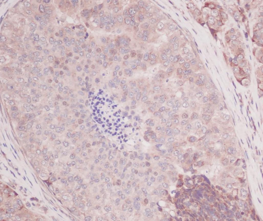
ARG56617 anti-CCL2 / MCP1 antibody IHC-P image
Immunohistochemistry: Formalin-fixed and paraffin-embedded sections of Human breast invasive ductal carcinoma. The recommended ARG56617 anti-CCL2 / MCP1 antibody concentration is 2.5 µg/ml and a two-hour incubation at RT. An HRP-labeled polymer detection system was used with a DAB chromogen. Antigen Retrieval: Boil tissue section in Sodium Citrate buffer (pH 6.0) followed by cooling at RT for 20 min.
-
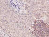
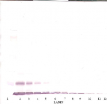
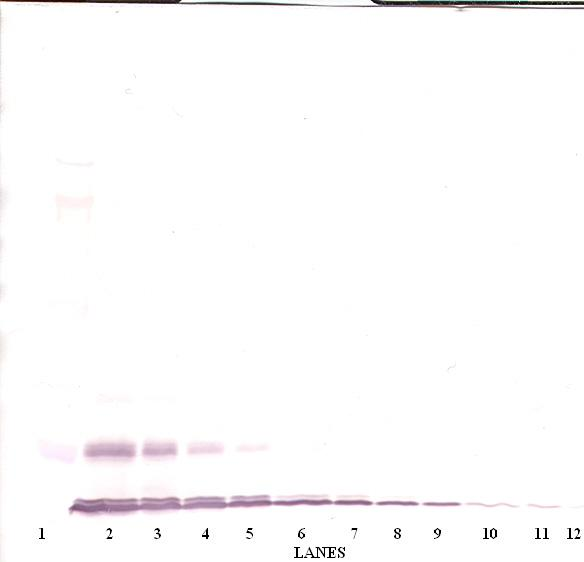
ARG56617 anti-CCL2 / MCP1 antibody WB image
Western blot: 250 - 0.24 ng of Human MCP-1(MCAF) stained with ARG56617 anti-CCL2 / MCP1 antibody, under non-reducing conditions.
-
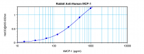
ARG56617 anti-CCL2 / MCP1 antibody standard curve image
Sandwich ELISA: ARG56617 anti-CCL2 / MCP1 antibody as a capture antibody at 0.5 - 2.0 µg/ml combined with ARG56731 anti-CCL2 / MCP1 antibody (Biotin) as a detection antibody. Results of a typical standard run with optical density.