ARG54724
anti-CCL2 / MCP1 antibody
anti-CCL2 / MCP1 antibody for Flow cytometry,ICC/IF,IHC-Formalin-fixed paraffin-embedded sections,Western blot and Human
Cancer antibody; Cell Biology and Cellular Response antibody; Immune System antibody; Metabolism antibody
Overview
| Product Description | Rabbit Polyclonal antibody recognizes CCL2 / MCP1 |
|---|---|
| Tested Reactivity | Hu |
| Predict Reactivity | Mk |
| Tested Application | FACS, ICC/IF, IHC-P, WB |
| Host | Rabbit |
| Clonality | Polyclonal |
| Isotype | IgG |
| Target Name | CCL2 / MCP1 |
| Antigen Species | Human |
| Immunogen | KLH-conjugated synthetic peptide corresponding to aa. 62-89 (C-terminus) of Human CCL2 (NP_002973.1). |
| Conjugation | Un-conjugated |
| Alternate Names | MCP1; Monocyte chemotactic and activating factor; MCAF; Monocyte chemotactic protein 1; Monocyte secretory protein JE; HSMCR30; Small-inducible cytokine A2; HC11; SMC-CF; GDCF-2; SCYA2; C-C motif chemokine 2; Monocyte chemoattractant protein 1; MCP-1 |
Application Instructions
| Application Suggestion |
|
||||||||||
|---|---|---|---|---|---|---|---|---|---|---|---|
| Application Note | * The dilutions indicate recommended starting dilutions and the optimal dilutions or concentrations should be determined by the scientist. | ||||||||||
| Positive Control | HeLa |
Properties
| Purification | This antibody is prepared by Saturated Ammonium Sulfate (SAS) precipitation followed by dialysis against PBS. |
|---|---|
| Buffer | PBS and 0.09% (W/V) Sodium azide |
| Preservative | 0.09% (W/V) Sodium azide |
| Storage Instruction | For continuous use, store undiluted antibody at 2-8°C for up to a week. For long-term storage, aliquot and store at -20°C or below. Storage in frost free freezers is not recommended. Avoid repeated freeze/thaw cycles. Suggest spin the vial prior to opening. The antibody solution should be gently mixed before use. |
| Note | For laboratory research only, not for drug, diagnostic or other use. |
Bioinformation
| Database Links | |
|---|---|
| Gene Symbol | CCL2 |
| Gene Full Name | chemokine (C-C motif) ligand 2 |
| Background | This gene is one of several cytokine genes clustered on the q-arm of chromosome 17. Chemokines are a superfamily of secreted proteins involved in immunoregulatory and inflammatory processes. The superfamily is divided into four subfamilies based on the arrangement of N-terminal cysteine residues of the mature peptide. This chemokine is a member of the CC subfamily which is characterized by two adjacent cysteine residues. This cytokine displays chemotactic activity for monocytes and basophils but not for neutrophils or eosinophils. It has been implicated in the pathogenesis of diseases characterized by monocytic infiltrates, like psoriasis, rheumatoid arthritis and atherosclerosis. It binds to chemokine receptors CCR2 and CCR4. [provided by RefSeq, Jul 2013] |
| Function | Chemotactic factor that attracts monocytes and basophils but not neutrophils or eosinophils. Augments monocyte anti-tumor activity. Has been implicated in the pathogenesis of diseases characterized by monocytic infiltrates, like psoriasis, rheumatoid arthritis or atherosclerosis. May be involved in the recruitment of monocytes into the arterial wall during the disease process of atherosclerosis. [From Uniprot] |
| Cellular Localization | Secreted. |
| Highlight | Related products: MCP1 antibodies; MCP1 ELISA Kits; MCP1 Duos / Panels; Related news: HMGB1 in inflammation Inflammatory Cytokines |
| Research Area | Cancer antibody; Cell Biology and Cellular Response antibody; Immune System antibody; Metabolism antibody |
| Calculated MW | 11 kDa |
| PTM | Processing at the N-terminus can regulate receptor and target cell selectivity. Deletion of the N-terminal residue converts it from an activator of basophil to an eosinophil chemoattractant. |
Images (4) Click the Picture to Zoom In
-
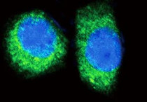
ARG54724 anti-CCL2 / MCP1 antibody ICC/IF image
Immunofluorescence: HeLa cells stained with ARG54724 anti-CCL2 / MCP1 antibody (green). DAPI (blue) for nuclear staining.
-
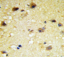
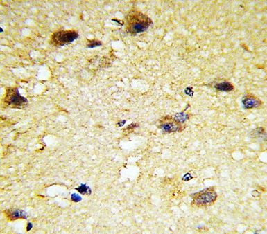
ARG54724 anti-CCL2 / MCP1 antibody IHC-P image
Immunohistochemistry: Paraffin-embedded Human brain tissue stained with ARG54724 anti-CCL2 / MCP1 antibody.
-

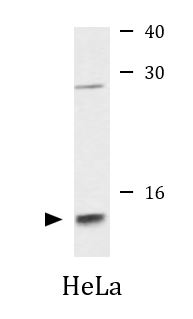
ARG54724 anti-CCL2 / MCP1 antibody WB image
Western blot: 35 µg of HeLa cell lysate stained with ARG54724 anti-CCL2 / MCP1 antibody.
-
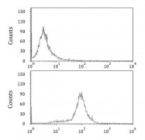
ARG54724 anti-CCL2 / MCP1 antibody FACS image
Flow Cytometry: HeLa cells stained with ARG54724 anti-CCL2 / MCP1 antibody (bottom histogram) or without primary antibody control (top histogram).