ARG41935
anti-C4 / Complement 4 antibody
anti-C4 / Complement 4 antibody for ICC/IF,Western blot and Human
Overview
| Product Description | Rabbit Polyclonal antibody recognizes C4 / Complement 4 |
|---|---|
| Tested Reactivity | Hu |
| Tested Application | ICC/IF, WB |
| Host | Rabbit |
| Clonality | Polyclonal |
| Isotype | IgG |
| Target Name | C4 / Complement 4 |
| Antigen Species | Human |
| Immunogen | Synthetic peptide of Human C4 / Complement 4. |
| Conjugation | Un-conjugated |
| Alternate Names | Complement C4-A; C4AD; CPAMD2; CO4; Acidic complement C4; C4S; C4A6; C4A4; RG; C4A2; C4A3; C3 and PZP-like alpha-2-macroglobulin domain-containing protein 2; C4 |
Application Instructions
| Application Suggestion |
|
||||||
|---|---|---|---|---|---|---|---|
| Application Note | * The dilutions indicate recommended starting dilutions and the optimal dilutions or concentrations should be determined by the scientist. | ||||||
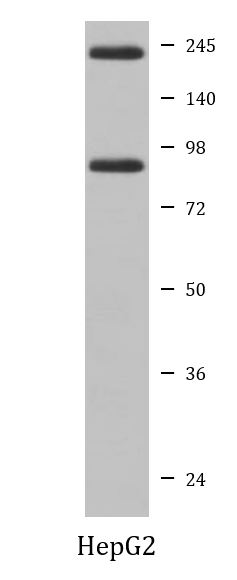
| Positive Control | HepG2 | ||||||
| Observed Size | ~ 210 kDa |
Properties
| Form | Liquid |
|---|---|
| Purification | Affinity purified. |
| Buffer | PBS (pH 7.4), 150 mM NaCl, 0.02% Sodium azide and 50% Glycerol. |
| Preservative | 0.02% Sodium azide |
| Stabilizer | 50% Glycerol |
| Storage Instruction | For continuous use, store undiluted antibody at 2-8°C for up to a week. For long-term storage, aliquot and store at -20°C. Storage in frost free freezers is not recommended. Avoid repeated freeze/thaw cycles. Suggest spin the vial prior to opening. The antibody solution should be gently mixed before use. |
| Note | For laboratory research only, not for drug, diagnostic or other use. |
Bioinformation
| Database Links | |
|---|---|
| Gene Symbol | C4A |
| Gene Full Name | complement component 4A (Rodgers blood group) |
| Background | This gene encodes the acidic form of complement factor 4, part of the classical activation pathway. The protein is expressed as a single chain precursor which is proteolytically cleaved into a trimer of alpha, beta, and gamma chains prior to secretion. The trimer provides a surface for interaction between the antigen-antibody complex and other complement components. The alpha chain is cleaved to release C4 anaphylatoxin, an antimicrobial peptide and a mediator of local inflammation. Deficiency of this protein is associated with systemic lupus erythematosus and type I diabetes mellitus. This gene localizes to the major histocompatibility complex (MHC) class III region on chromosome 6. Varying haplotypes of this gene cluster exist, such that individuals may have 1, 2, or 3 copies of this gene. Two transcript variants encoding different isoforms have been found for this gene. [provided by RefSeq, Nov 2014] |
| Function | Non-enzymatic component of C3 and C5 convertases and thus essential for the propagation of the classical complement pathway. Covalently binds to immunoglobulins and immune complexes and enhances the solubilization of immune aggregates and the clearance of IC through CR1 on erythrocytes. C4A isotype is responsible for effective binding to form amide bonds with immune aggregates or protein antigens, while C4B isotype catalyzes the transacylation of the thioester carbonyl group to form ester bonds with carbohydrate antigens. Derived from proteolytic degradation of complement C4, C4a anaphylatoxin is a mediator of local inflammatory process. It induces the contraction of smooth muscle, increases vascular permeability and causes histamine release from mast cells and basophilic leukocytes. [UniProt] |
| Cellular Localization | Secreted. Cell junction, synapse. Cell projection, axon. Cell projection, dendrite. [UniProt] |
| Calculated MW | 193 kDa |
| PTM | Prior to secretion, the single-chain precursor is enzymatically cleaved to yield non-identical chains alpha, beta and gamma. During activation, the alpha chain is cleaved by C1 into C4a and C4b, and C4b stays linked to the beta and gamma chains. Further degradation of C4b by C1 into the inactive fragments C4c and C4d blocks the generation of C3 convertase. The proteolytic cleavages often are incomplete so that many structural forms can be found in plasma. N- and O-glycosylated. O-glycosylated with a core 1 or possibly core 8 glycan. [UniProt] |
Images (1) Click the Picture to Zoom In
-

ARG41935 anti-C4 / Complement 4 antibody WB image
Western blot: HepG2 cell lysate stained with ARG41935 anti-C4 / Complement 4 antibody.