ARG40995
anti-C1QBP antibody
anti-C1QBP antibody for Flow cytometry,ICC/IF,IHC-Formalin-fixed paraffin-embedded sections,Western blot and Human,Mouse,Rat
Overview
| Product Description | Rabbit Polyclonal antibody recognizes C1QBP |
|---|---|
| Tested Reactivity | Hu, Ms, Rat |
| Tested Application | FACS, ICC/IF, IHC-P, WB |
| Host | Rabbit |
| Clonality | Polyclonal |
| Isotype | IgG |
| Target Name | C1QBP |
| Antigen Species | Human |
| Immunogen | Recombinant protein corresponding to E190-Q282 of Human C1QBP. |
| Conjugation | Un-conjugated |
| Alternate Names | SF2p32; ASF/SF2-associated protein p32; gC1Q-R; C1qBP; gC1qR; GC1QBP; gC1q-R protein; HABP1; Mitochondrial matrix protein p32; p32; p33; Complement component 1 Q subcomponent-binding protein, mitochondrial; Glycoprotein gC1qBP; Hyaluronan-binding protein 1 |
Application Instructions
| Application Suggestion |
|
||||||||||
|---|---|---|---|---|---|---|---|---|---|---|---|
| Application Note | IHC-P: Antigen Retrieval: Heat mediation was performed in Citrate buffer (pH 6.0, epitope retrieval solution) for 20 min. * The dilutions indicate recommended starting dilutions and the optimal dilutions or concentrations should be determined by the scientist. |
Properties
| Form | Liquid |
|---|---|
| Purification | Affinity purification with immunogen. |
| Buffer | 0.2% Na2HPO4, 0.9% NaCl, 0.05% Sodium azide and 5% BSA. |
| Preservative | 0.05% Sodium azide |
| Stabilizer | 5% BSA |
| Concentration | 0.5 mg/ml |
| Storage Instruction | For continuous use, store undiluted antibody at 2-8°C for up to a week. For long-term storage, aliquot and store at -20°C or below. Storage in frost free freezers is not recommended. Avoid repeated freeze/thaw cycles. Suggest spin the vial prior to opening. The antibody solution should be gently mixed before use. |
| Note | For laboratory research only, not for drug, diagnostic or other use. |
Bioinformation
| Database Links |
Swiss-port # O35796 Rat Complement component 1 Q subcomponent-binding protein, mitochondrial Swiss-port # Q07021 Human Complement component 1 Q subcomponent-binding protein, mitochondrial |
|---|---|
| Gene Symbol | C1QBP |
| Gene Full Name | complement component 1, q subcomponent binding protein |
| Background | The human complement subcomponent C1q associates with C1r and C1s in order to yield the first component of the serum complement system. The protein encoded by this gene is known to bind to the globular heads of C1q molecules and inhibit C1 activation. This protein has also been identified as the p32 subunit of pre-mRNA splicing factor SF2, as well as a hyaluronic acid-binding protein. [provided by RefSeq, Jul 2008] |
| Function | Is believed to be a multifunctional and multicompartmental protein involved in inflammation and infection processes, ribosome biogenesis, regulation of apoptosis, transcriptional regulation and pre-mRNA splicing. At the cell surface is thought to act as an endothelial receptor for plasma proteins of the complement and kallikrein-kinin cascades. Putative receptor for C1q; specifically binds to the globular "heads" of C1q thus inhibiting C1; may perform the receptor function through a complex with C1qR/CD93. In complex with cytokeratin-1/KRT1 is a high affinity receptor for kininogen-1/HMWK. Can also bind other plasma proteins, such as coagulation factor XII leading to its autoactivation. May function to bind initially fluid kininogen-1 to the cell membrane. The secreted form may enhance both extrinsic and intrinsic coagulation pathways. It is postulated that the cell surface form requires docking with transmembrane proteins for downstream signaling which might be specific for a cell-type or response. By acting as C1q receptor is involved in chemotaxis of immature dendritic cells and neutrophils and is proposed to signal through CD209/DC-SIGN on immature dendritic cells, through integrin alpha-4/beta-1 during trophoblast invasion of the decidua, and through integrin beta-1 during endothelial cell adhesion and spreading. Signaling involved in inhibition of innate immune response is implicating the PI3K-AKT/PKB pathway. In mitochondrial translation may be involved in formation of functional 55S mitoribosomes; the function seems to involve its RNA-binding activity. May be involved in the nucleolar ribosome maturation process; the function may involve the exchange of FBL for RRP1 in the association with pre-ribosome particles. Involved in regulation of RNA splicing by inhibiting the RNA-binding capacity of SRSF1 and its phosphorylation. Is required for the nuclear translocation of splicing factor U2AF1L4. Involved in regulation of CDKN2A- and HRK-mediated apoptosis. Stabilizes mitochondrial CDKN2A isoform smARF. May be involved in regulation of FOXC1 transcriptional activity and NFY/CCAAT-binding factor complex-mediated transcription. In infection processes acts as an attachment site for microbial proteins, including Listeria monocytogenes internalin B and Staphylococcus aureus protein A. May play a role in antibacterial defense as it can bind to cell surface hyaluronan and inhibit Streptococcus pneumoniae hyaluronate lyase. Involved in replication of Rubella virus. May be involved in modulation of the immune response; ligation by HCV core protein is resulting in suppresion of interleukin-12 production in monocyte-derived dendritic cells. Involved in regulation of antiviral response by inhibiting DDX58- and IFIH1-mediated signaling pathways probably involving its association with MAVS after viral infection. Involved in HIV-1 replication, presumably by contributing to splicing of viral RNA. [UniProt] |
| Cellular Localization | Mitochondrion matrix. Nucleus. Cell membrane; Peripheral membrane protein; Extracellular side. Secreted. Cytoplasm. Nucleus, nucleolus. Note=Seems to be predominantly localized to mitochondria. Secreted by activated lymphocytes. [UniProt] |
| Calculated MW | 31 kDa |
Images (6) Click the Picture to Zoom In
-
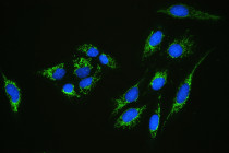
ARG40995 anti-C1QBP antibody ICC/IF image
Immunofluorescence: U2OS cells stained with ARG40995 anti-C1QBP antibody (green) at 2 µg/ml, overnight at 4°C. DAPI (blue) for nuclear staining.
-
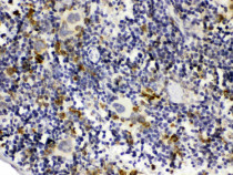
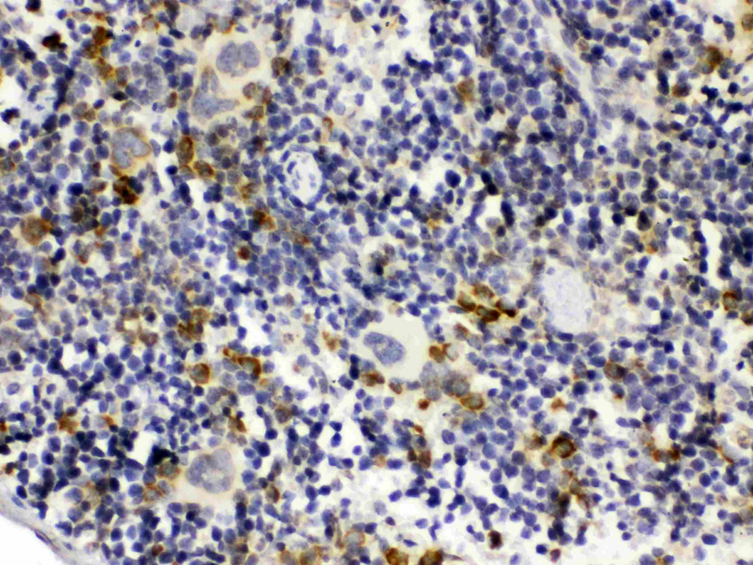
ARG40995 anti-C1QBP antibody IHC-P image
Immunohistochemistry: Paraffin-embedded Mouse spleen tissue. Antigen Retrieval: Heat mediation was performed in Citrate buffer (pH 6.0, epitope retrieval solution) for 20 min. The tissue section was blocked with 10% goat serum. The tissue section was then stained with ARG40995 anti-C1QBP antibody at 1 µg/ml dilution, overnight at 4°C.
-
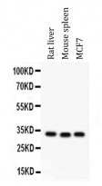
ARG40995 anti-C1QBP antibody WB image
Western blot: 50 µg of samples under reducing conditions. Rat liver, Mouse spleen and MCF7 whole cell lysates stained with ARG40995 anti-C1QBP antibody at 0.5 µg/ml, overnight at 4°C.
-
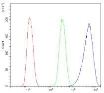
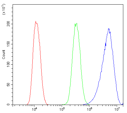
ARG40995 anti-C1QBP antibody FACS image
Flow Cytometry: U2OS cells were blocked with 10% normal goat serum and then stained with ARG40995 anti-C1QBP antibody (blue) at 1 µg/10^6 cells for 30 min at 20°C, followed by incubation with DyLight®488 labelled secondary antibody. Isotype control antibody (green) was Rabbit IgG (1 µg/10^6 cells) used under the same conditions. Unlabelled sample (red) was also used as a control.
-
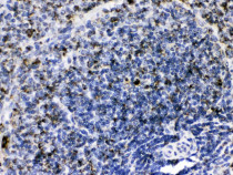
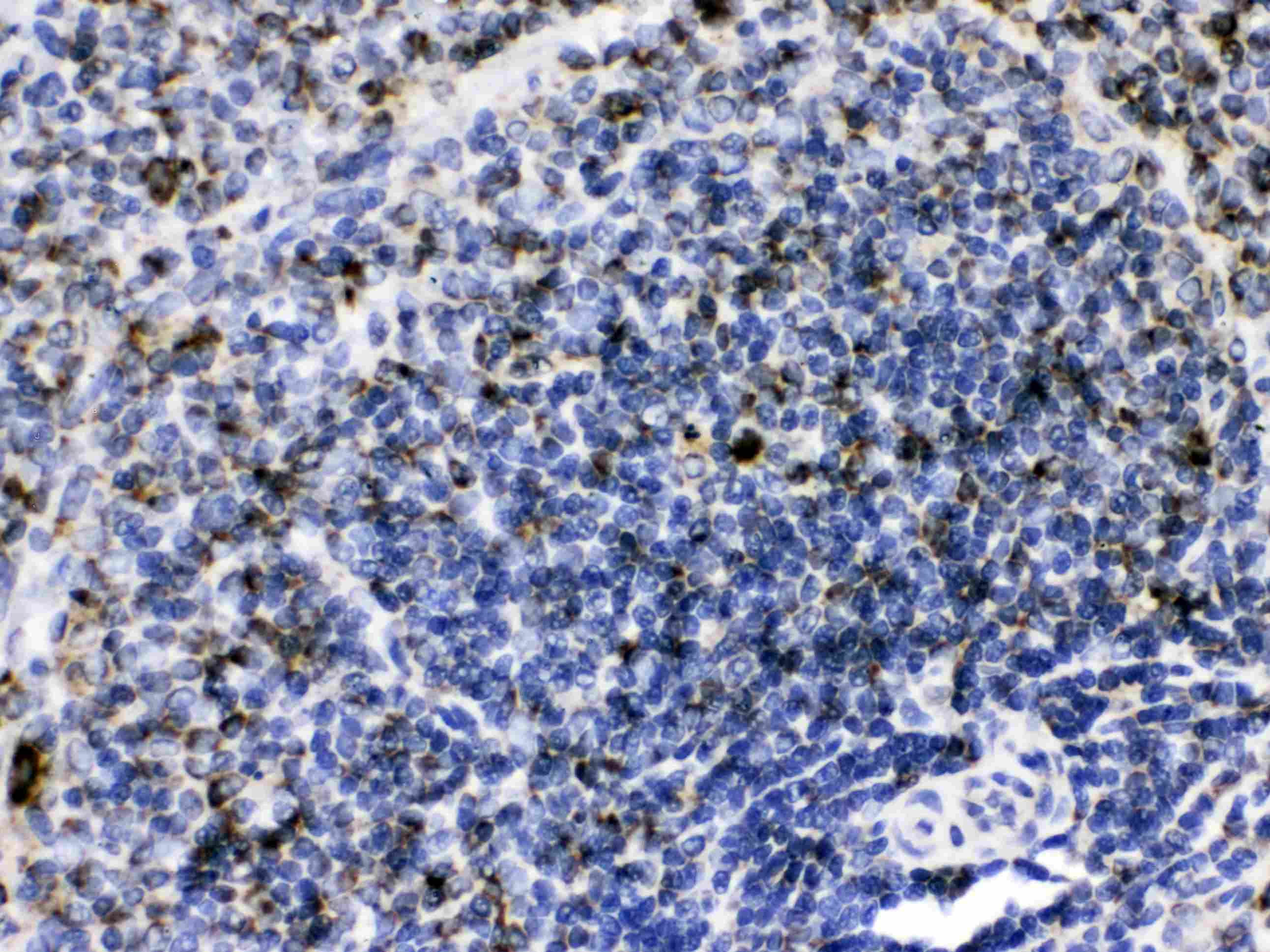
ARG40995 anti-C1QBP antibody IHC-P image
Immunohistochemistry: Paraffin-embedded Rat spleen tissue. Antigen Retrieval: Heat mediation was performed in Citrate buffer (pH 6.0, epitope retrieval solution) for 20 min. The tissue section was blocked with 10% goat serum. The tissue section was then stained with ARG40995 anti-C1QBP antibody at 1 µg/ml dilution, overnight at 4°C.
-

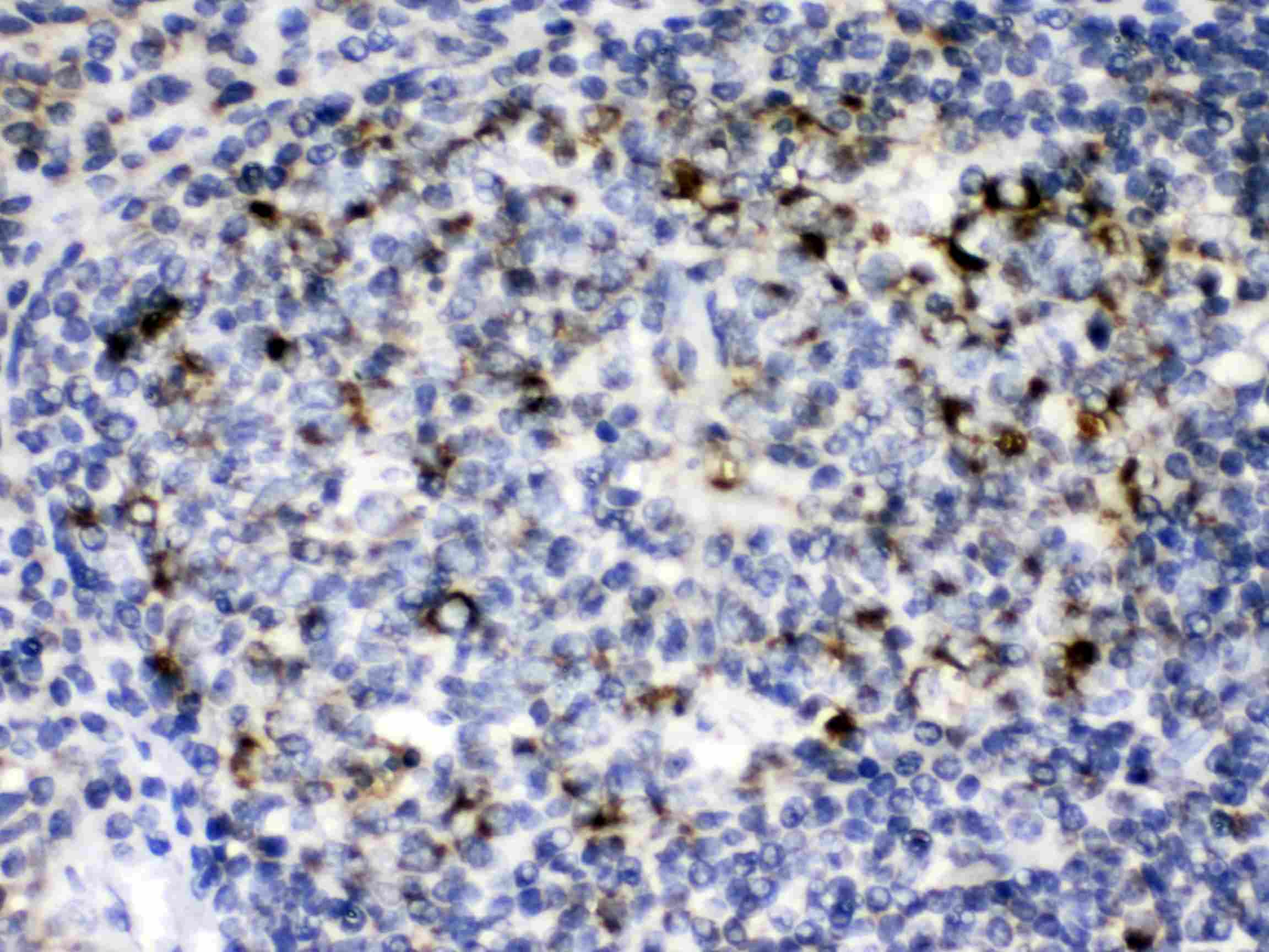
ARG40995 anti-C1QBP antibody IHC-P image
Immunohistochemistry: Paraffin-embedded Human tonsil tissue. Antigen Retrieval: Heat mediation was performed in Citrate buffer (pH 6.0, epitope retrieval solution) for 20 min. The tissue section was blocked with 10% goat serum. The tissue section was then stained with ARG40995 anti-C1QBP antibody at 1 µg/ml dilution, overnight at 4°C.