ARG41381
anti-BubR1 antibody
anti-BubR1 antibody for Flow cytometry,IHC-Formalin-fixed paraffin-embedded sections,Immunoprecipitation,Western blot and Human
Overview
| Product Description | Rabbit Polyclonal antibody recognizes BubR1 |
|---|---|
| Tested Reactivity | Hu |
| Tested Application | FACS, IHC-P, IP, WB |
| Host | Rabbit |
| Clonality | Polyclonal |
| Isotype | IgG |
| Target Name | BubR1 |
| Antigen Species | Human |
| Immunogen | Synthetic peptide derived from Human BubR1. |
| Conjugation | Un-conjugated |
| Alternate Names | MVA1; Bub1A; Mitotic checkpoint serine/threonine-protein kinase BUB1 beta; MAD3/BUB1-related protein kinase; SSK1; Mitotic checkpoint kinase MAD3L; Protein SSK1; hBUBR1; EC 2.7.11.1; MAD3L; BUBR1; BUB1beta |
Application Instructions
| Application Suggestion |
|
||||||||||
|---|---|---|---|---|---|---|---|---|---|---|---|
| Application Note | * The dilutions indicate recommended starting dilutions and the optimal dilutions or concentrations should be determined by the scientist. | ||||||||||
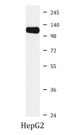
| Observed Size | ~ 120 kDa |
Properties
| Form | Liquid |
|---|---|
| Purification | Affinity purified. |
| Buffer | PBS (pH 7.4), 150 mM NaCl, 0.02% Sodium azide and 50% Glycerol. |
| Preservative | 0.02% Sodium azide |
| Stabilizer | 50% Glycerol |
| Storage Instruction | For continuous use, store undiluted antibody at 2-8°C for up to a week. For long-term storage, aliquot and store at -20°C. Storage in frost free freezers is not recommended. Avoid repeated freeze/thaw cycles. Suggest spin the vial prior to opening. The antibody solution should be gently mixed before use. |
| Note | For laboratory research only, not for drug, diagnostic or other use. |
Bioinformation
| Database Links |
Swiss-port # O60566 Human Mitotic checkpoint serine/threonine-protein kinase BUB1 beta |
|---|---|
| Gene Symbol | BUB1B |
| Gene Full Name | BUB1 mitotic checkpoint serine/threonine kinase B |
| Background | This gene encodes a kinase involved in spindle checkpoint function. The protein has been localized to the kinetochore and plays a role in the inhibition of the anaphase-promoting complex/cyclosome (APC/C), delaying the onset of anaphase and ensuring proper chromosome segregation. Impaired spindle checkpoint function has been found in many forms of cancer. [provided by RefSeq, Jul 2008] |
| Function | Essential component of the mitotic checkpoint. Required for normal mitosis progression. The mitotic checkpoint delays anaphase until all chromosomes are properly attached to the mitotic spindle. One of its checkpoint functions is to inhibit the activity of the anaphase-promoting complex/cyclosome (APC/C) by blocking the binding of CDC20 to APC/C, independently of its kinase activity. The other is to monitor kinetochore activities that depend on the kinetochore motor CENPE. Required for kinetochore localization of CENPE. Negatively regulates PLK1 activity in interphase cells and suppresses centrosome amplification. Also implicated in triggering apoptosis in polyploid cells that exit aberrantly from mitotic arrest. May play a role for tumor suppression. [UniProt] |
| Cellular Localization | Cytoplasm. Nucleus. Chromosome, centromere, kinetochore. Cytoplasm, cytoskeleton, microtubule organizing center, centrosome. Note=Cytoplasmic in interphase cells. Associates with the kinetochores in early prophase. Kinetochore localization requires BUB1, PLK1 and KNL1. [UniProt] |
| Calculated MW | 120 kDa |
| PTM | Proteolytically cleaved by caspase-3 in a cell cycle specific manner. The cleavage might be involved in the durability of the cell cycle delay. Caspase-3 cleavage is associated with abrogation of the mitotic checkpoint. The major site of cleavage is at Asp-610. Acetylation at Lys-250 regulates its degradation and timing in anaphase entry. Ubiquitinated. Degraded by the proteasome. Sumoylated with SUMO2 and SUMO3. The sumoylation mediates the association with CENPE at the kinetochore. Autophosphorylated in vitro. Intramolecular autophosphorylation is stimulated by CENPE. Phosphorylated during mitosis and hyperphosphorylated in mitotically arrested cells. Phosphorylation at Ser-670 and Ser-1043 occurs at kinetochores upon mitotic entry with dephosphorylation at the onset of anaphase. [UniProt] |
Images (1) Click the Picture to Zoom In
-
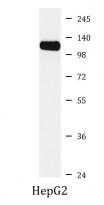
ARG41381 anti-BubR1 antibody WB image
Western blot: HepG2 cell lysate stained with ARG41381 anti-BubR1 antibody.