ARG10775
anti-Bestrophin 3 antibody
anti-Bestrophin 3 antibody for Confocal microscopy (Confocal),Dot blot,ELISA,ICC/IF,IHC-Formalin-fixed paraffin-embedded sections,Immunoprecipitation,Western blot and Mouse,Rat
Overview
| Product Description | Rabbit Polyclonal antibody recognizes Bestrophin 3 |
|---|---|
| Tested Reactivity | Ms, Rat |
| Tested Application | Confocal, Dot, ELISA, ICC/IF, IHC-P, IP, WB |
| Host | Rabbit |
| Clonality | Polyclonal |
| Isotype | IgG |
| Target Name | Bestrophin 3 |
| Antigen Species | Rat |
| Immunogen | Synthetic peptide taken within the last 50 aa (C-terminus) from Rat Bestrophin 3. |
| Conjugation | Un-conjugated |
| Alternate Names | VMD2L3; Bestrophin-3; Vitelliform macular dystrophy 2-like protein 3 |
Application Instructions
| Application Suggestion |
|
||||||||||||||||
|---|---|---|---|---|---|---|---|---|---|---|---|---|---|---|---|---|---|
| Application Note | * The dilutions indicate recommended starting dilutions and the optimal dilutions or concentrations should be determined by the scientist. |
Properties
| Form | Liquid |
|---|---|
| Purification | Affinity purified. |
| Buffer | Tris-Glycine Buffer (pH 7.4 - 7.8), Hepes, 0.02% Sodium azide, 30% Glycerol and 0.5% BSA. |
| Preservative | 0.02% Sodium azide |
| Stabilizer | 30% Glycerol and 0.5% BSA |
| Concentration | 0.5 mg/ml |
| Storage Instruction | For continuous use, store undiluted antibody at 2-8°C for up to a week. For long-term storage, aliquot and store at -20°C. Storage in frost free freezers is not recommended. Avoid repeated freeze/thaw cycles. Suggest spin the vial prior to opening. The antibody solution should be gently mixed before use. |
| Note | For laboratory research only, not for drug, diagnostic or other use. |
Bioinformation
| Database Links | |
|---|---|
| Gene Symbol | Best3 |
| Gene Full Name | bestrophin 3 |
| Background | BEST3 belongs to the bestrophin family of anion channels, which includes BEST1 (MIM 607854), the gene mutant in vitelliform macular dystrophy (VMD; MIM 153700), and 2 other BEST1-like genes, BEST2 (MIM 607335) and BEST4 (MIM 607336). Bestrophins are transmembrane (TM) proteins that share a homology region containing a high content of aromatic residues, including an invariant arg-phe-pro (RFP) motif. The bestrophin genes share a conserved gene structure, with almost identical sizes of the 8 RFP-TM domain-encoding exons and highly conserved exon-intron boundaries. Each of the 4 bestrophin genes has a unique 3-prime end of variable length (Stohr et al., 2002 [PubMed 12032738]; Tsunenari et al., 2003 [PubMed 12907679]).[supplied by OMIM, Mar 2008] |
| Function | Forms calcium-sensitive chloride channels. Permeable to bicarbonate. [UniProt] |
| Calculated MW | 76 kDa |
Images (1) Click the Picture to Zoom In
-
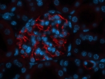
ARG10775 anti-Bestrophin 3 antibody IHC-P image
Immunohistochemistry: PFA-fixed and paraffin-embedded Mouse glomerulus stained with ARG10775 anti-Bestrophin 3 antibody at 1:100 dilution. Incubation for 36 h at 4°C. Requires antigen retrieval.


