ARG40576
anti-Bcl XL antibody
anti-Bcl XL antibody for Flow cytometry,ICC/IF,IHC-Formalin-fixed paraffin-embedded sections,Immunoprecipitation,Western blot and Human,Mouse,Rat
Overview
| Product Description | Rabbit Polyclonal antibody recognizes Bcl XL |
|---|---|
| Tested Reactivity | Hu, Ms, Rat |
| Tested Application | FACS, ICC/IF, IHC-P, IP, WB |
| Host | Rabbit |
| Clonality | Polyclonal |
| Isotype | IgG |
| Target Name | Bcl XL |
| Antigen Species | Human |
| Immunogen | Synthetic peptide derived from Human Bcl XL. |
| Conjugation | Un-conjugated |
| Alternate Names | Apoptosis regulator Bcl-X; BCLXS; BCL-XL/S; PPP1R52; bcl-xS; Bcl-2-like protein 1; Bcl2-L-1; Bcl-X; BCLX; bcl-xL; BCL2L; BCLXL |
Application Instructions
| Application Suggestion |
|
||||||||||||
|---|---|---|---|---|---|---|---|---|---|---|---|---|---|
| Application Note | * The dilutions indicate recommended starting dilutions and the optimal dilutions or concentrations should be determined by the scientist. |
Properties
| Form | Liquid |
|---|---|
| Purification | Affinity purified. |
| Buffer | PBS (pH 7.4), 150 mM NaCl, 0.02% Sodium azide and 50% Glycerol. |
| Preservative | 0.02% Sodium azide |
| Stabilizer | 50% Glycerol |
| Storage Instruction | For continuous use, store undiluted antibody at 2-8°C for up to a week. For long-term storage, aliquot and store at -20°C. Storage in frost free freezers is not recommended. Avoid repeated freeze/thaw cycles. Suggest spin the vial prior to opening. The antibody solution should be gently mixed before use. |
| Note | For laboratory research only, not for drug, diagnostic or other use. |
Bioinformation
| Database Links | |
|---|---|
| Gene Symbol | BCL2L1 |
| Gene Full Name | BCL2-like 1 |
| Background | The protein encoded by this gene belongs to the BCL-2 protein family. BCL-2 family members form hetero- or homodimers and act as anti- or pro-apoptotic regulators that are involved in a wide variety of cellular activities. The proteins encoded by this gene are located at the outer mitochondrial membrane, and have been shown to regulate outer mitochondrial membrane channel (VDAC) opening. VDAC regulates mitochondrial membrane potential, and thus controls the production of reactive oxygen species and release of cytochrome C by mitochondria, both of which are the potent inducers of cell apoptosis. Two alternatively spliced transcript variants, which encode distinct isoforms, have been reported. The longer isoform acts as an apoptotic inhibitor and the shorter form acts as an apoptotic activator. [provided by RefSeq, Jul 2008] |
| Function | Potent inhibitor of cell death. Inhibits activation of caspases. Appears to regulate cell death by blocking the voltage-dependent anion channel (VDAC) by binding to it and preventing the release of the caspase activator, CYC1, from the mitochondrial membrane. Also acts as a regulator of G2 checkpoint and progression to cytokinesis during mitosis. Isoform Bcl-X(L) also regulates presynaptic plasticity, including neurotransmitter release and recovery, number of axonal mitochondria as well as size and number of synaptic vesicle clusters. During synaptic stimulation, increases ATP availability from mitochondria through regulation of mitochondrial membrane ATP synthase F(1)F(0) activity and regulates endocytic vesicle retrieval in hippocampal neurons through association with DMN1L and stimulation of its GTPase activity in synaptic vesicles. Isoform Bcl-X(S) promotes apoptosis. [UniProt] |
| Cellular Localization | Isoform Bcl-X(L): Mitochondrion inner/outer membrane. Mitochondrion matrix. Cytoplasmic vesicle, secretory vesicle, synaptic vesicle membrane. Cytoplasm, cytosol, cytoskeleton, microtubule organizing center, centrosome. Nucleus membrane; Single-pass membrane protein; Cytoplasmic side. Note=After neuronal stimulation, translocates from cytosol to synaptic vesicle and mitochondrion membrane in a calmodulin-dependent manner. Localizes to the centrosome when phosphorylated at Ser-49. [UniProt] |
| Calculated MW | 26 kDa |
| PTM | Proteolytically cleaved by caspases during apoptosis. The cleaved protein, lacking the BH4 motif, has pro-apoptotic activity. Phosphorylated on Ser-62 by CDK1. This phosphorylation is partial in normal mitotic cells, but complete in G2-arrested cells upon DNA-damage, thus promoting subsequent apoptosis probably by triggering caspases-mediated proteolysis. Phosphorylated by PLK3, leading to regulate the G2 checkpoint and progression to cytokinesis during mitosis. Phosphorylation at Ser-49 appears during the S phase and G2, disappears rapidly in early mitosis during prometaphase, metaphase and early anaphase, and re-appears during telophase and cytokinesis. [UniProt] |
Images (3) Click the Picture to Zoom In
-
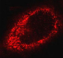
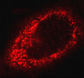
ARG40576 anti-Bcl XL antibody ICC/IF image
Immunofluorescence: HeLa cells stained with ARG40576 anti-Bcl XL antibody.
-
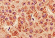
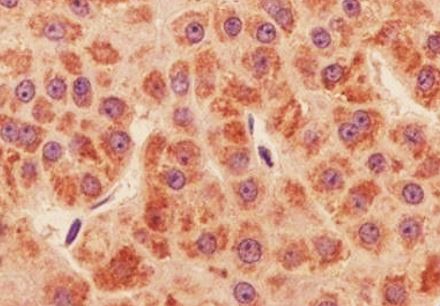
ARG40576 anti-Bcl XL antibody IHC-P image
Immunohistochemistry: Paraffin-embedded Human liver cancer tissue stained with ARG40576 anti-Bcl XL antibody.
-
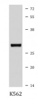
ARG40576 anti-Bcl XL antibody WB image
Western blot: K562 cell lysate stained with ARG40576 anti-Bcl XL antibody.