ARG54147
anti-Bax antibody
anti-Bax antibody for Western blot and Human,Monkey
Cancer antibody; Cell Biology and Cellular Response antibody; Cell Death antibody; Metabolism antibody; Mitochondrial fission antibody; Apoptosis Marker antibody; Pro-apoptotic Bcl2 protein antibody
Overview
| Product Description | Mouse Monoclonal antibody recognizes BAX |
|---|---|
| Tested Reactivity | Hu, Mk |
| Tested Application | WB |
| Host | Mouse |
| Clonality | Monoclonal |
| Isotype | IgG2a |
| Target Name | Bax |
| Antigen Species | Human |
| Immunogen | Purified recombinant human Bax protein fragments expressed in E.coli |
| Conjugation | Un-conjugated |
| Alternate Names | Bcl-2-like protein 4; Bcl2-L-4; BCL2L4; Apoptosis regulator BAX |
Application Instructions
| Application Suggestion |
|
||||
|---|---|---|---|---|---|
| Application Note | * The dilutions indicate recommended starting dilutions and the optimal dilutions or concentrations should be determined by the scientist. |
Properties
| Form | Liquid |
|---|---|
| Purification | Affinity purified |
| Buffer | PBS (pH 7.4), 0.02% Sodium azide and 50% Glycerol |
| Preservative | 0.02% Sodium azide |
| Stabilizer | 50% Glycerol |
| Concentration | 5.5 mg/ml |
| Storage Instruction | For continuous use, store undiluted antibody at 2-8°C for up to a week. For long-term storage, aliquot and store at -20°C. Storage in frost free freezers is not recommended. Avoid repeated freeze/thaw cycles. Suggest spin the vial prior to opening. The antibody solution should be gently mixed before use. |
| Note | For laboratory research only, not for drug, diagnostic or other use. |
Bioinformation
| Database Links | |
|---|---|
| Gene Symbol | BAX |
| Gene Full Name | BCL2-associated X protein |
| Background | Bax belongs to the BCL2 protein family. BCL2 family members form hetero- or homodimers and act as anti- or pro-apoptotic regulators that are involved in a wide variety of cellular activities. This protein forms a heterodimer with BCL2, and functions as an apoptotic activator. The association and the ratio of BAX to BCL2 also determines survival or death of a cell following an apoptotic stimulus. This protein is reported to interact with, and increase the opening of, the mitochondrial voltage-dependent anion channel (VDAC), which leads to the loss in membrane potential and the release of cytochrome c. The expression of this gene is regulated by the tumor suppressor P53 and has been shown to be involved in P53-mediated apoptosis. Multiple alternatively spliced transcript variants, which encode different isoforms, have been reported for this gene. [provided by RefSeq, Dec 2019] |
| Function | Bax plays a role in the mitochondrial apoptotic process. Under normal conditions, BAX is largely cytosolic via constant retrotranslocation from mitochondria to the cytosol mediated by BCL2L1/Bcl-xL, which avoids accumulation of toxic BAX levels at the mitochondrial outer membrane (MOM) (PubMed:21458670). Under stress conditions, undergoes a conformation change that causes translocation to the mitochondrion membrane, leading to the release of cytochrome c that then triggers apoptosis. Promotes activation of CASP3, and thereby apoptosis. [UniProt] |
| Cellular Localization | Isoform Alpha: Mitochondrion membrane; Single-pass membrane protein. Cytoplasm. |
| Research Area | Cancer antibody; Cell Biology and Cellular Response antibody; Cell Death antibody; Metabolism antibody; Mitochondrial fission antibody; Apoptosis Marker antibody; Pro-apoptotic Bcl2 protein antibody |
| Calculated MW | 21 kDa |
Images (1) Click the Picture to Zoom In
-
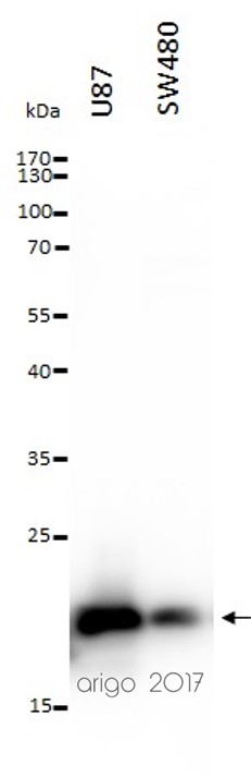
ARG54147 anti-Bax antibody WB image
Western blot: 20 µl of U87 and SW480 cell lysates stained with ARG54147 anti-Bax antibody at 1:1000 dilution.