ARG54681
anti-Bad antibody
anti-Bad antibody for ELISA,ICC/IF,IHC-Formalin-fixed paraffin-embedded sections,Western blot and Human,Mouse,Rat
Cancer antibody; Cell Biology and Cellular Response antibody; Cell Death antibody; Metabolism antibody

Overview
| Product Description | Rabbit Polyclonal antibody recognizes Bad |
|---|---|
| Tested Reactivity | Hu, Ms, Rat |
| Tested Application | ELISA, ICC/IF, IHC-P, WB |
| Host | Rabbit |
| Clonality | Polyclonal |
| Isotype | IgG |
| Target Name | Bad |
| Immunogen | Synthetic peptide (15 aa) within aa. 90-140 of Human Bad protein. |
| Conjugation | Un-conjugated |
| Alternate Names | Bcl-2-binding component 6; Bcl-2-like protein 8; BCL2L8; Bcl-xL/Bcl-2-associated death promoter; BAD; Bcl2-associated agonist of cell death; BBC2; Bcl2 antagonist of cell death; Bcl2-L-8 |
Application Instructions
| Application Suggestion |
|
||||||||||
|---|---|---|---|---|---|---|---|---|---|---|---|
| Application Note | * The dilutions indicate recommended starting dilutions and the optimal dilutions or concentrations should be determined by the scientist. | ||||||||||
| Positive Control | T24 Cell Lysate |
Properties
| Form | Liquid |
|---|---|
| Purification | Ion exchange chromatography. |
| Buffer | PBS and 0.02% Sodium azide |
| Preservative | 0.02% Sodium azide |
| Concentration | 1 mg/ml |
| Storage Instruction | For continuous use, store undiluted antibody at 2-8°C for up to a week. For long-term storage, aliquot and store at -20°C or below. Storage in frost free freezers is not recommended. Avoid repeated freeze/thaw cycles. Suggest spin the vial prior to opening. The antibody solution should be gently mixed before use. |
| Note | For laboratory research only, not for drug, diagnostic or other use. |
Bioinformation
| Database Links | |
|---|---|
| Gene Symbol | BAD |
| Gene Full Name | BCL2-associated agonist of cell death |
| Background | Members in the Bcl-2 family are critical regulators of apoptosis by either inhibiting or promoting cell death. Bcl-2 homology 3 (BH3) domain containing pro-apoptotic proteins, such as Bax, Bid, and Bik, form a growing subclass of the Bcl-2 family. Another such protein is the Bcl-2-antagonist of cell death (Bad). Bad regulates apoptosis by forming heterodimers with anti-apoptotic proteins Bcl-2 and Bcl-xL, thereby preventing them from binding with Bax. Bad activity is regulated by its phosphorylation; it is inactivated by kinases such as Akt and MAP kinase and thus promotes cell survival, whereas JNK-induced phosphorylation promotes the apoptotic role of Bad. |
| Function | Promotes cell death. Successfully competes for the binding to Bcl-X(L), Bcl-2 and Bcl-W, thereby affecting the level of heterodimerization of these proteins with BAX. Can reverse the death repressor activity of Bcl-X(L), but not that of Bcl-2 (By similarity). Appears to act as a link between growth factor receptor signaling and the apoptotic pathways. [UniProt] |
| Research Area | Cancer antibody; Cell Biology and Cellular Response antibody; Cell Death antibody; Metabolism antibody |
| Calculated MW | 18 kDa |
| PTM | Phosphorylated on one or more of Ser-75, Ser-99, Ser-118 and Ser-134 in response to survival stimuli, which blocks its pro-apoptotic activity. Phosphorylation on Ser-99 or Ser-75 promotes heterodimerization with 14-3-3 proteins. This interaction then facilitates the phosphorylation at Ser-118, a site within the BH3 motif, leading to the release of Bcl-X(L) and the promotion of cell survival. Ser-99 is the major site of AKT/PKB phosphorylation, Ser-118 the major site of protein kinase A (CAPK) phosphorylation. Phosphorylation at Ser-99 by PKB/AKT1 is almost completely blocked by the apoptotic C-terminus cleavage product of PKN2 generated by caspases-3 activity during apoptosis. Methylation at Arg-94 and Arg-96 by PRMT1 inhibits Akt-mediated phosphorylation at Ser-99. |
Images (3) Click the Picture to Zoom In
-
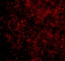
ARG54681 anti-BAD antibody ICC/IF image
Immunofluorescence: rat thymus cells stained with ARG54681 anti-BAD antibody at 10 μg/ml.
-

ARG54681 anti-BAD antibody IHC image
Immunohistochemical: rat thymus stained with ARG54681 anti-Bad at 2 μg/ml.
-
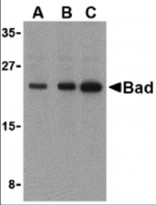
ARG54681 anti-BAD antibody WB image
Western blot: T24 cell lysates stained with ARG54681 anti-Bad antibody at (A) 0.5, (B) 1, and (C) 2 μg/ml.


Customer's Feedback
Average
Review for anti-Bad antibody
Application:WB
Sample:Mouse ovary
Sample Loading Amount:30 µg
Primary Antibody Dilution Factor:1:500
Primary Antibody Incubation Time:overnight
Primary Antibody Incubation Temperature:4 ºC

Average

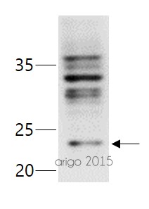
Review for anti-Bad antibody
Application:WB
Sample:PC3
Sample Loading Amount:30 µg
Primary Antibody Dilution Factor:1:500
Primary Antibody Incubation Time:overnight
Primary Antibody Incubation Temperature:4 ºC


