ARG40585
anti-Bad antibody
anti-Bad antibody for Flow cytometry,ICC/IF,IHC-Formalin-fixed paraffin-embedded sections,Immunoprecipitation,Western blot and Human,Mouse,Rat
Overview
| Product Description | Rabbit Polyclonal antibody recognizes Bad |
|---|---|
| Tested Reactivity | Hu, Ms, Rat |
| Tested Application | FACS, ICC/IF, IHC-P, IP, WB |
| Host | Rabbit |
| Clonality | Polyclonal |
| Isotype | IgG |
| Target Name | Bad |
| Antigen Species | Human |
| Immunogen | Synthetic peptide derived from Human Bad. |
| Conjugation | Un-conjugated |
| Alternate Names | Bcl-2-binding component 6; Bcl-2-like protein 8; BCL2L8; Bcl-xL/Bcl-2-associated death promoter; BAD; Bcl2-associated agonist of cell death; BBC2; Bcl2 antagonist of cell death; Bcl2-L-8 |
Application Instructions
| Application Suggestion |
|
||||||||||||
|---|---|---|---|---|---|---|---|---|---|---|---|---|---|
| Application Note | * The dilutions indicate recommended starting dilutions and the optimal dilutions or concentrations should be determined by the scientist. |
Properties
| Form | Liquid |
|---|---|
| Purification | Affinity purified. |
| Buffer | PBS (pH 7.4), 150 mM NaCl, 0.02% Sodium azide and 50% Glycerol. |
| Preservative | 0.02% Sodium azide |
| Stabilizer | 50% Glycerol |
| Storage Instruction | For continuous use, store undiluted antibody at 2-8°C for up to a week. For long-term storage, aliquot and store at -20°C. Storage in frost free freezers is not recommended. Avoid repeated freeze/thaw cycles. Suggest spin the vial prior to opening. The antibody solution should be gently mixed before use. |
| Note | For laboratory research only, not for drug, diagnostic or other use. |
Bioinformation
| Database Links | |
|---|---|
| Gene Symbol | BAD |
| Gene Full Name | BCL2-associated agonist of cell death |
| Background | The protein encoded by this gene is a member of the BCL-2 family. BCL-2 family members are known to be regulators of programmed cell death. This protein positively regulates cell apoptosis by forming heterodimers with BCL-xL and BCL-2, and reversing their death repressor activity. Proapoptotic activity of this protein is regulated through its phosphorylation. Protein kinases AKT and MAP kinase, as well as protein phosphatase calcineurin were found to be involved in the regulation of this protein. Alternative splicing of this gene results in two transcript variants which encode the same isoform. [provided by RefSeq, Jul 2008] |
| Function | Promotes cell death. Successfully competes for the binding to Bcl-X(L), Bcl-2 and Bcl-W, thereby affecting the level of heterodimerization of these proteins with BAX. Can reverse the death repressor activity of Bcl-X(L), but not that of Bcl-2 (By similarity). Appears to act as a link between growth factor receptor signaling and the apoptotic pathways. [UniProt] |
| Cellular Localization | Mitochondrion outer membrane. Cytoplasm. Note=Colocalizes with HIF3A in the cytoplasm (By similarity). Upon phosphorylation, locates to the cytoplasm. [UniProt] |
| Calculated MW | 18 kDa |
| PTM | Phosphorylated on one or more of Ser-75, Ser-99, Ser-118 and Ser-134 in response to survival stimuli, which blocks its pro-apoptotic activity. Phosphorylation on Ser-99 or Ser-75 promotes heterodimerization with 14-3-3 proteins. This interaction then facilitates the phosphorylation at Ser-118, a site within the BH3 motif, leading to the release of Bcl-X(L) and the promotion of cell survival. Ser-99 is the major site of AKT/PKB phosphorylation, Ser-118 the major site of protein kinase A (CAPK) phosphorylation. Phosphorylation at Ser-99 by PKB/AKT1 is almost completely blocked by the apoptotic C-terminus cleavage product of PKN2 generated by caspases-3 activity during apoptosis. Methylation at Arg-94 and Arg-96 by PRMT1 inhibits Akt-mediated phosphorylation at Ser-99. [UniProt] |
Images (3) Click the Picture to Zoom In
-
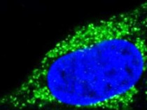
ARG40585 anti-Bad antibody ICC/IF image
Immunofluorescence: HeLa cells stained with ARG40585 anti-Bad antibody.
-
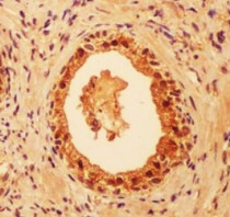
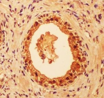
ARG40585 anti-Bad antibody IHC-P image
Immunohistochemistry: Paraffin-embedded Human colon cancer tissue stained with ARG40585 anti-Bad antibody.
-

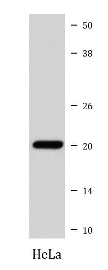
ARG40585 anti-Bad antibody WB image
Western blot: HeLa cell lysate stained with ARG40585 anti-Bad antibody.