ARG54832
anti-BMPR2 antibody
anti-BMPR2 antibody for Flow cytometry,IHC-Formalin-fixed paraffin-embedded sections,Western blot and Human,Mouse
Cell Biology and Cellular Response antibody; Developmental Biology antibody; Signaling Transduction antibody
Overview
| Product Description | Rabbit Polyclonal antibody recognizes BMPR2 |
|---|---|
| Tested Reactivity | Hu, Ms |
| Tested Application | FACS, IHC-P, WB |
| Host | Rabbit |
| Clonality | Polyclonal |
| Isotype | IgG |
| Target Name | BMPR2 |
| Antigen Species | Human |
| Immunogen | KLH-conjugated synthetic peptide corresponding to aa. 28-59 (N-terminus) of Human BMPR2. |
| Conjugation | Un-conjugated |
| Alternate Names | T-ALK; BMPR-II; EC 2.7.11.30; PPH1; BRK-3; BMR2; Bone morphogenetic protein receptor type II; Bone morphogenetic protein receptor type-2; BMPR3; BMP type II receptor; POVD1; BMPR-2; BMP type-2 receptor |
Application Instructions
| Application Suggestion |
|
||||||||
|---|---|---|---|---|---|---|---|---|---|
| Application Note | * The dilutions indicate recommended starting dilutions and the optimal dilutions or concentrations should be determined by the scientist. | ||||||||
| Positive Control | Mouse heart |
Properties
| Form | Liquid |
|---|---|
| Purification | Purification with Protein G. |
| Buffer | PBS and 0.09% (W/V) Sodium azide |
| Preservative | 0.09% (W/V) Sodium azide |
| Storage Instruction | For continuous use, store undiluted antibody at 2-8°C for up to a week. For long-term storage, aliquot and store at -20°C or below. Storage in frost free freezers is not recommended. Avoid repeated freeze/thaw cycles. Suggest spin the vial prior to opening. The antibody solution should be gently mixed before use. |
| Note | For laboratory research only, not for drug, diagnostic or other use. |
Bioinformation
| Database Links |
Swiss-port # O35607 Mouse Bone morphogenetic protein receptor type-2 Swiss-port # Q13873 Human Bone morphogenetic protein receptor type-2 |
|---|---|
| Gene Symbol | BMPR2 |
| Gene Full Name | bone morphogenetic protein receptor, type II (serine/threonine kinase) |
| Background | This gene encodes a member of the bone morphogenetic protein (BMP) receptor family of transmembrane serine/threonine kinases. The ligands of this receptor are BMPs, which are members of the TGF-beta superfamily. BMPs are involved in endochondral bone formation and embryogenesis. These proteins transduce their signals through the formation of heteromeric complexes of two different types of serine (threonine) kinase receptors: type I receptors of about 50-55 kD and type II receptors of about 70-80 kD. Type II receptors bind ligands in the absence of type I receptors, but they require their respective type I receptors for signaling, whereas type I receptors require their respective type II receptors for ligand binding. Mutations in this gene have been associated with primary pulmonary hypertension, both familial and fenfluramine-associated, and with pulmonary venoocclusive disease. [provided by RefSeq, Jul 2008] |
| Function | On ligand binding, forms a receptor complex consisting of two type II and two type I transmembrane serine/threonine kinases. Type II receptors phosphorylate and activate type I receptors which autophosphorylate, then bind and activate SMAD transcriptional regulators. Binds to BMP-7, BMP-2 and, less efficiently, BMP-4. Binding is weak but enhanced by the presence of type I receptors for BMPs. [UniProt] |
| Cellular Localization | Membrane; Single-pass type I membrane protein |
| Research Area | Cell Biology and Cellular Response antibody; Developmental Biology antibody; Signaling Transduction antibody |
| Calculated MW | 115 kDa |
Images (3) Click the Picture to Zoom In
-
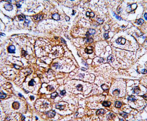
ARG54832 anti-BMPR2 antibody IHC-P image
Immunohistochemistry: Formalin-fixed and paraffin-embedded Human hepatocarcinoma tissue stained with ARG54832 anti-BMPR2 antibody.
-
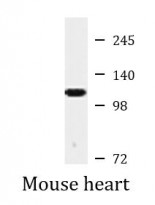
ARG54832 anti-BMPR2 antibody WB image
Western blot: Mouse heart lysate stained with ARG54832 anti-BMPR2 antibody.
-
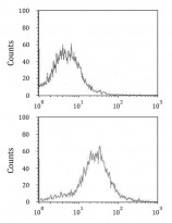
ARG54832 anti-BMPR2 antibody FACS image
Flow Cytometry: HepG2 cells stained with ARG54832 anti-BMPR2 antibody (bottom histogram) or without primary antibody control (top histogram), followed by incubation with FITC labelled secondary antibody.


