ARG63184
anti-BLNK / SLP65 antibody
anti-BLNK / SLP65 antibody for Flow cytometry,ICC/IF,Western blot and Human
Immune System antibody; Signaling Transduction antibody
Overview
| Product Description | Goat Polyclonal antibody recognizes BLNK / SLP65 |
|---|---|
| Tested Reactivity | Hu |
| Predict Reactivity | Ms, Rat, Cow, Dog, Pig |
| Tested Application | FACS, ICC/IF, WB |
| Specificity | This antibody is expected to recognize both reported isoforms (NP_037446.1; NP_001107566.1). |
| Host | Goat |
| Clonality | Polyclonal |
| Isotype | IgG |
| Target Name | BLNK / SLP65 |
| Antigen Species | Human |
| Immunogen | C-KDSTRLKYAVKVS |
| Conjugation | Un-conjugated |
| Alternate Names | SLP65; BLNK-S; B-cell linker protein; bca; B-cell adapter containing a Src homology 2 domain protein; SLP-65; AGM4; LY57; Cytoplasmic adapter protein; Src homology 2 domain-containing leukocyte protein of 65 kDa; BASH; B-cell adapter containing a SH2 domain protein |
Application Instructions
| Application Suggestion |
|
||||||||
|---|---|---|---|---|---|---|---|---|---|
| Application Note | WB: Recommend incubate at RT for 1h. * The dilutions indicate recommended starting dilutions and the optimal dilutions or concentrations should be determined by the scientist. |
Properties
| Form | Liquid |
|---|---|
| Purification | Purified from goat serum by antigen affinity chromatography. |
| Buffer | Tris saline (pH 7.3), 0.02% Sodium azide and 0.5% BSA. |
| Preservative | 0.02% Sodium azide |
| Stabilizer | 0.5% BSA |
| Concentration | 0.5 mg/ml |
| Storage Instruction | For continuous use, store undiluted antibody at 2-8°C for up to a week. For long-term storage, aliquot and store at -20°C or below. Storage in frost free freezers is not recommended. Avoid repeated freeze/thaw cycles. Suggest spin the vial prior to opening. The antibody solution should be gently mixed before use. |
| Note | For laboratory research only, not for drug, diagnostic or other use. |
Bioinformation
| Database Links | |
|---|---|
| Background | This gene encodes a cytoplasmic linker or adaptor protein that plays a critical role in B cell development. This protein bridges B cell receptor-associated kinase activation with downstream signaling pathways, thereby affecting various biological functions. The phosphorylation of five tyrosine residues is necessary for this protein to nucleate distinct signaling effectors following B cell receptor activation. Mutations in this gene cause hypoglobulinemia and absent B cells, a disease in which the pro- to pre-B-cell transition is developmentally blocked. Deficiency in this protein has also been shown in some cases of pre-B acute lymphoblastic leukemia. Alternatively spliced transcript variants have been found for this gene. [provided by RefSeq, May 2012] |
| Research Area | Immune System antibody; Signaling Transduction antibody |
| Calculated MW | 50 kDa |
| PTM | Following BCR activation, phosphorylated on tyrosine residues by SYK and LYN. When phosphorylated, serves as a scaffold to assemble downstream targets of antigen activation, including PLCG1, VAV1, GRB2 and NCK1. Phosphorylation of Tyr-84, Tyr-178 and Tyr-189 facilitates PLCG1 binding. Phosphorylation of Tyr-96 facilitates BTK binding. Phosphorylation of Tyr-72 facilitates VAV1 and NCK1 binding. Phosphorylation is required for both Ca(2+) and MAPK signaling pathways. |
Images (4) Click the Picture to Zoom In
-

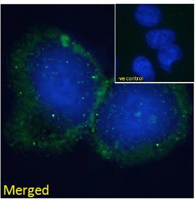
ARG63184 anti-BLNK / SLP65 antibody ICC/IF image
Immunofluorescence: Paraformaldehyde fixed HepG2 cells permeabilized with 0.15% Triton. Cells were stained with ARG63184 anti-BLNK / SLP65 antibody (green) at 10 µg/ml dilution for 1 hour. DAPI (blue) for nuclear staining. Negative control: Unimmunized goat IgG (green) at 10 µg/ml dilution.
-
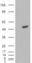
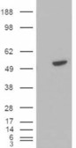
ARG63184 anti-BLNK / SLP65 antibody WB image
Western blot: 1). Mock transfection; 2) BLNK (RC202488) expressing plasmid transfected HEK293 cell lysate standed with ARG63184 anti-BLNK / SLP65 antibody.
-

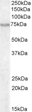
ARG63184 anti-BLNK / SLP65 antibody WB image
Western blot: 35 µg of Daudi cell lysate (in RIPA buffer) stained with ARG63184 anti-BLNK / SLP65 antibody at 0.3 µg/ml dilution and incubated at RT for 1 hour.
-
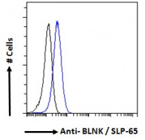
ARG63184 anti-BLNK / SLP65 antibody FACS image
Flow Cytometry: Paraformaldehyde-fixed Daudi cells permeabilized with 0.5% Triton. Cells were stained with ARG63184 anti-BLNK / SLP65 antibody (blue line) at 10 µg/ml dilution for 1 hour, followed by incubation with Alexa FluorR 488 labelled secondary antibody. IgG control: Unimmunized goat IgG (black line).