ARG58209
anti-BIRC2 / cIAP1 antibody
anti-BIRC2 / cIAP1 antibody for Flow cytometry,ICC/IF,Western blot and Human,Mouse,Rat
Overview
| Product Description | Rabbit Polyclonal antibody recognizes BIRC2 / cIAP1 |
|---|---|
| Tested Reactivity | Hu, Ms, Rat |
| Tested Application | FACS, ICC/IF, WB |
| Host | Rabbit |
| Clonality | Polyclonal |
| Isotype | IgG |
| Target Name | BIRC2 / cIAP1 |
| Antigen Species | Human |
| Immunogen | E. coli-derived Human cIAP1 recombinant protein (Position: D320-T570). |
| Conjugation | Un-conjugated |
| Alternate Names | BIRC2; Baculoviral IAP Repeat Containing 2; C-IAP1; RNF48; MIHB; Hiap-2; CIAP1; API1; Baculoviral IAP Repeat-Containing Protein 2; RING-Type E3 Ubiquitin Transferase BIRC2; TNFR2-TRAF-Signaling Complex Protein 2; NFR2-TRAF Signalling Complex Protein; Cellular Inhibitor Of Apoptosis 1; Inhibitor Of Apoptosis Protein 2; RING Finger Protein 48; Apoptosis Inhibitor 1; IAP Homolog B; HIAP2; Baculoviral IAP Repeat-Containing 2; EC 2.3.2.27; HIAP-2; IAP-2 |
Application Instructions
| Application Suggestion |
|
||||||||
|---|---|---|---|---|---|---|---|---|---|
| Application Note | * The dilutions indicate recommended starting dilutions and the optimal dilutions or concentrations should be determined by the scientist. |
Properties
| Form | Liquid |
|---|---|
| Buffer | 0.9% NaCl, 0.2% Na2HPO4, 0.05% Sodium azide and 4% Trehalose. |
| Preservative | 0.05% Sodium azide |
| Stabilizer | 4% Trehalose |
| Concentration | 0.5 mg/ml |
| Storage Instruction | For continuous use, store undiluted antibody at 2-8°C for up to a week. For long-term storage, aliquot and store at -20°C or below. Storage in frost free freezers is not recommended. Avoid repeated freeze/thaw cycles. Suggest spin the vial prior to opening. The antibody solution should be gently mixed before use. |
| Note | For laboratory research only, not for drug, diagnostic or other use. |
Bioinformation
| Database Links |
Swiss-port # Q13490 Human Baculoviral IAP repeat-containing protein 2 Swiss-port # Q62210 Mouse Baculoviral IAP repeat-containing protein 2 |
|---|---|
| Gene Symbol | BIRC2 |
| Gene Full Name | baculoviral IAP repeat containing 2 |
| Background | The protein encoded by this gene is a member of a family of proteins that inhibits apoptosis by binding to tumor necrosis factor receptor-associated factors TRAF1 and TRAF2, probably by interfering with activation of ICE-like proteases. This encoded protein inhibits apoptosis induced by serum deprivation and menadione, a potent inducer of free radicals. Alternatively spliced transcript variants encoding different isoforms have been found for this gene. [provided by RefSeq, Jan 2012] |
| Function | Multi-functional protein which regulates not only caspases and apoptosis, but also modulates inflammatory signaling and immunity, mitogenic kinase signaling, and cell proliferation, as well as cell invasion and metastasis. Acts as an E3 ubiquitin-protein ligase regulating NF-kappa-B signaling and regulates both canonical and non-canonical NF-kappa-B signaling by acting in opposite directions: acts as a positive regulator of the canonical pathway and suppresses constitutive activation of non-canonical NF-kappa-B signaling. The target proteins for its E3 ubiquitin-protein ligase activity include: RIPK1, RIPK2, RIPK3, RIPK4, CASP3, CASP7, CASP8, TRAF2, DIABLO/SMAC, MAP3K14/NIK, MAP3K5/ASK1, IKBKG/NEMO, IKBKE and MXD1/MAD1. Can also function as an E3 ubiquitin-protein ligase of the NEDD8 conjugation pathway, targeting effector caspases for neddylation and inactivation. Acts as an important regulator of innate immune signaling via regulation of Toll-like receptors (TLRs), Nodlike receptors (NLRs) and RIG-I like receptors (RLRs), collectively referred to as pattern recognition receptors (PRRs). Protects cells from spontaneous formation of the ripoptosome, a large multi-protein complex that has the capability to kill cancer cells in a caspase-dependent and caspase-independent manner. Suppresses ripoptosome formation by ubiquitinating RIPK1 and CASP8. Can stimulate the transcriptional activity of E2F1. Plays a role in the modulation of the cell cycle. |
| Cellular Localization | Cytoplasm; Nucleus. [UniProt] |
| Calculated MW | 70 kDa |
| PTM | Ubl conjugation. [UniProt] |
Images (3) Click the Picture to Zoom In
-

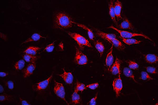
ARG58209 anti-BIRC2 / cIAP1 antibody ICC/IF image
Immunofluorescence: U2OS cells were blocked with 10% goat serum and then stained with ARG58209 anti-BIRC2 / cIAP1 antibody (red) at 2 µg/ml dilution, overnight at 4°C. DAPI (blue) for nuclear staining.
-
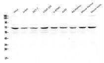
ARG58209 anti-BIRC2 / cIAP1 antibody WB image
Western blot: 50 µg of HeLa whole cell lysates, Jurkat whole cell lysates, MCF-7 whole cell lysates, COLO-320 whole cell lysates, U-87MG whole cell lysates, A549 whole cell lysates, Rat thymus, Mouse thymus and Mouse testis lysates stained with ARG58209 anti-BIRC2 / cIAP1 antibody at 0.5 µg/ml, overnight at 4°C, under reducing conditions.
-
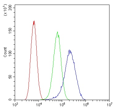
ARG58209 anti-BIRC2 / cIAP1 antibody FACS image
Flow Cytometry: U2OS cells were blocked with 10% normal goat serum and then stained with ARG58209 anti-BIRC2 / cIAP1 antibody (blue) at 1 µg/10^6 cells for 30 min at 20°C, followed by incubation with DyLight®488 labelled secondary antibody. Isotype control antibody (green) was rabbit IgG (1 µg/10^6 cells) used under the same conditions. Unlabelled sample (red) was also used as a control.