ARG43090
anti-BAG6 / BAT3 antibody
anti-BAG6 / BAT3 antibody for Flow cytometry,ICC/IF,IHC-Formalin-fixed paraffin-embedded sections,Western blot and Human,Mouse,Rat
Overview
| Product Description | Rabbit Polyclonal antibody recognizes BAG6 / BAT3 |
|---|---|
| Tested Reactivity | Hu, Ms, Rat |
| Tested Application | FACS, ICC/IF, IHC-P, WB |
| Host | Rabbit |
| Clonality | Polyclonal |
| Isotype | IgG |
| Target Name | BAG6 / BAT3 |
| Antigen Species | Human |
| Immunogen | Recombinant protein corresponding to D15-A88 of Human BAG6 / BAT3. |
| Conjugation | Un-conjugated |
| Alternate Names | BAG-6; G3; BAG6; Protein G3; BCL2-associated athanogene 6; Protein Scythe; BAT3; HLA-B-associated transcript 3; BAG family molecular chaperone regulator 6; D6S52E; Large proline-rich protein BAG6 |
Application Instructions
| Application Suggestion |
|
||||||||||
|---|---|---|---|---|---|---|---|---|---|---|---|
| Application Note | IHC-P: Antigen Retrieval: Heat mediation was performed in Citrate buffer (pH 6.0) for 20 min. * The dilutions indicate recommended starting dilutions and the optimal dilutions or concentrations should be determined by the scientist. |
||||||||||
| Observed Size | ~ 160 kDa |
Properties
| Form | Liquid |
|---|---|
| Purification | Affinity purification with immunogen. |
| Buffer | 0.2% Na2HPO4, 0.9% NaCl, 0.05% Sodium azide and 4% Trehalose. |
| Preservative | 0.05% Sodium azide |
| Stabilizer | 4% Trehalose |
| Concentration | 0.5 mg/ml |
| Storage Instruction | For continuous use, store undiluted antibody at 2-8°C for up to a week. For long-term storage, aliquot and store at -20°C or below. Storage in frost free freezers is not recommended. Avoid repeated freeze/thaw cycles. Suggest spin the vial prior to opening. The antibody solution should be gently mixed before use. |
| Note | For laboratory research only, not for drug, diagnostic or other use. |
Bioinformation
| Database Links | |
|---|---|
| Gene Symbol | BAG6 |
| Gene Full Name | BCL2-associated athanogene 6 |
| Background | This gene was first characterized as part of a cluster of genes located within the human major histocompatibility complex class III region. This gene encodes a nuclear protein that is cleaved by caspase 3 and is implicated in the control of apoptosis. In addition, the protein forms a complex with E1A binding protein p300 and is required for the acetylation of p53 in response to DNA damage. Multiple transcript variants encoding different isoforms have been found for this gene. [provided by RefSeq, Jul 2008] |
| Function | ATP-independent molecular chaperone preventing the aggregation of misfolded and hydrophobic patches-containing proteins (PubMed:21636303). Functions as part of a cytosolic protein quality control complex, the BAG6/BAT3 complex, which maintains these client proteins in a soluble state and participates to their proper delivery to the endoplasmic reticulum or alternatively can promote their sorting to the proteasome where they undergo degradation (PubMed:20516149, PubMed:21636303, PubMed:21743475, PubMed:28104892). The BAG6/BAT3 complex is involved in the post-translational delivery of tail-anchored/type II transmembrane proteins to the endoplasmic reticulum membrane. Recruited to ribosomes, it interacts with the transmembrane region of newly synthesized tail-anchored proteins and together with SGTA and ASNA1 mediates their delivery to the endoplasmic reticulum (PubMed:20516149, PubMed:20676083, PubMed:28104892, PubMed:25535373). Client proteins that cannot be properly delivered to the endoplasmic reticulum are ubiquitinated by RNF126, an E3 ubiquitin-protein ligase associated with BAG6 and are sorted to the proteasome (PubMed:24981174, PubMed:28104892, PubMed:27193484). SGTA which prevents the recruitment of RNF126 to BAG6 may negatively regulate the ubiquitination and the proteasomal degradation of client proteins (PubMed:23129660, PubMed:25179605, PubMed:27193484). Similarly, the BAG6/BAT3 complex also functions as a sorting platform for proteins of the secretory pathway that are mislocalized to the cytosol either delivering them to the proteasome for degradation or to the endoplasmic reticulum (PubMed:21743475). The BAG6/BAT3 complex also plays a role in the endoplasmic reticulum-associated degradation (ERAD), a quality control mechanism that eliminates unwanted proteins of the endoplasmic reticulum through their retrotranslocation to the cytosol and their targeting to the proteasome. It maintains these retrotranslocated proteins in an unfolded yet soluble state condition in the cytosol to ensure their proper delivery to the proteasome (PubMed:21636303). BAG6 is also required for selective ubiquitin-mediated degradation of defective nascent chain polypeptides by the proteasome. In this context, it may participate to the production of antigenic peptides and play a role in antigen presentation in immune response (By similarity). BAG6 is also involved in endoplasmic reticulum stress-induced pre-emptive quality control, a mechanism that selectively attenuates the translocation of newly synthesized proteins into the endoplasmic reticulum and reroutes them to the cytosol for proteasomal degradation. BAG6 may ensure the proper degradation of these proteins and thereby protects the endoplasmic reticulum from protein overload upon stress (PubMed:26565908). By inhibiting the polyubiquitination and subsequent proteasomal degradation of HSPA2 it may also play a role in the assembly of the synaptonemal complex during spermatogenesis (By similarity). Also positively regulates apoptosis by interacting with and stabilizing the proapoptotic factor AIFM1 (By similarity). By controlling the steady-state expression of the IGF1R receptor, indirectly regulates the insulin-like growth factor receptor signaling pathway (PubMed:26692333). Involved in DNA damage-induced apoptosis: following DNA damage, accumulates in the nucleus and forms a complex with p300/EP300, enhancing p300/EP300-mediated p53/TP53 acetylation leading to increase p53/TP53 transcriptional activity (PubMed:17403783). When nuclear, may also act as a component of some chromatin regulator complex that regulates histone 3 'Lys-4' dimethylation (H3K4me2) (PubMed:18765639). Released extracellularly via exosomes, it is a ligand of the natural killer/NK cells receptor NCR3 and stimulates NK cells cytotoxicity. It may thereby trigger NK cells cytotoxicity against neighboring tumor cells and immature myeloid dendritic cells (DC). Mediates ricin-induced apoptosis. [UniProt] |
| Cellular Localization | Cytoplasm, cytosol. Nucleus. Secreted, exosome. Note=Normally localized in cytosol and nucleus, it can also be released extracellularly, in exosomes, by tumor and myeloid dendritic cells. [UniProt] |
| Calculated MW | 119 kDa |
| PTM | Ricin can induce a cleavage by the caspase CASP3. The released C-terminal peptide induces apoptosis. (Microbial infection) In case of infection by L.pneumophila, ubiquitinated by the SCF(LegU1) complex. [UniProt] |
Images (15) Click the Picture to Zoom In
-
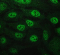
ARG43090 anti-BAG6 / BAT3 antibody ICC/IF image
Immunofluorescence: U2OS cells were blocked with 10% goat serum and then stained with ARG43090 anti-BAG6 / BAT3 antibody at 2 µg/ml dilution, overnight at 4°C.
-

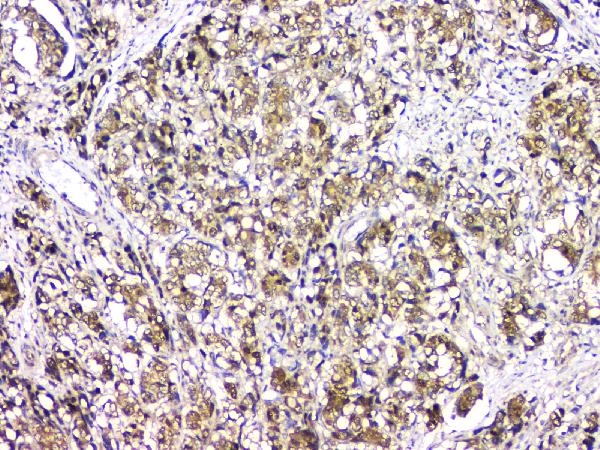
ARG43090 anti-BAG6 / BAT3 antibody IHC-P image
Immunohistochemistry: Paraffin-embedded Human gastric cancer tissue. Antigen Retrieval: Heat mediation was performed in Citrate buffer (pH 6.0) for 20 min. The tissue section was blocked with 10% goat serum. The tissue section was then stained with ARG43090 anti-BAG6 / BAT3 antibody at 1 µg/ml dilution, overnight at 4°C.
-
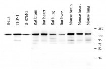
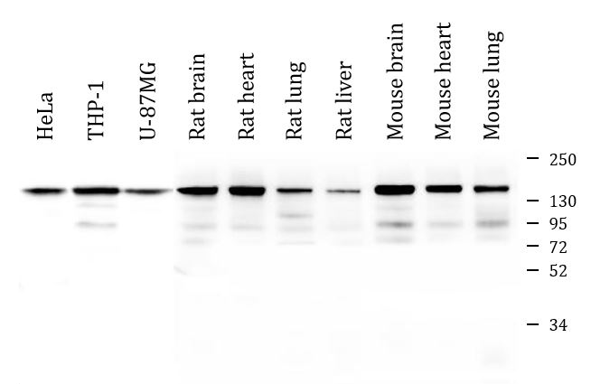
ARG43090 anti-BAG6 / BAT3 antibody WB image
Western blot: 50 µg of sample under reducing conditions. HeLa, THP-1, U-87MG, Rat brain, Rat heart, Rat lung, Rat liver, Mouse brain, Mouse heart and Mouse lung lysates stained with ARG43090 anti-BAG6 / BAT3 antibody at 0.5 µg/ml dilution, overnight at 4°C.
-

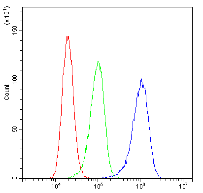
ARG43090 anti-BAG6 / BAT3 antibody FACS image
Flow Cytometry: A431 cells were blocked with 10% normal goat serum and then stained with ARG43090 anti-BAG6 / BAT3 antibody (blue) at 1 µg/10^6 cells for 30 min at 20°C, followed by incubation with DyLight®488 labelled secondary antibody. Isotype control antibody (green) was rabbit IgG (1 µg/10^6 cells) used under the same conditions. Unlabelled sample (red) was also used as a control.
-
ARG43090 anti-BAG6 / BAT3 antibody IHC-P image
Immunohistochemistry: Paraffin-embedded Human intestinal cancer tissue. Antigen Retrieval: Heat mediation was performed in Citrate buffer (pH 6.0) for 20 min. The tissue section was blocked with 10% goat serum. The tissue section was then stained with ARG43090 anti-BAG6 / BAT3 antibody at 1 µg/ml dilution, overnight at 4°C.
-
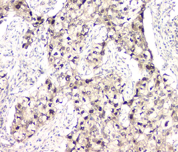
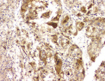
ARG43090 anti-BAG6 / BAT3 antibody IHC-P image
Immunohistochemistry: Paraffin-embedded Human lung cancer tissue. Antigen Retrieval: Heat mediation was performed in Citrate buffer (pH 6.0) for 20 min. The tissue section was blocked with 10% goat serum. The tissue section was then stained with ARG43090 anti-BAG6 / BAT3 antibody at 1 µg/ml dilution, overnight at 4°C.
-
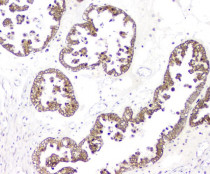
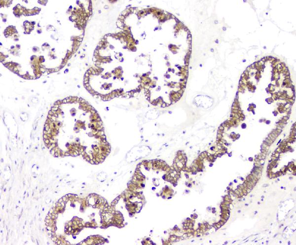
ARG43090 anti-BAG6 / BAT3 antibody IHC-P image
Immunohistochemistry: Paraffin-embedded Human ovary cancer tissue. Antigen Retrieval: Heat mediation was performed in Citrate buffer (pH 6.0) for 20 min. The tissue section was blocked with 10% goat serum. The tissue section was then stained with ARG43090 anti-BAG6 / BAT3 antibody at 1 µg/ml dilution, overnight at 4°C.
-
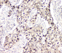
ARG43090 anti-BAG6 / BAT3 antibody IHC-P image
Immunohistochemistry: Paraffin-embedded Human liver cancer tissue. Antigen Retrieval: Heat mediation was performed in Citrate buffer (pH 6.0) for 20 min. The tissue section was blocked with 10% goat serum. The tissue section was then stained with ARG43090 anti-BAG6 / BAT3 antibody at 1 µg/ml dilution, overnight at 4°C.
-
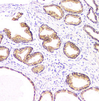
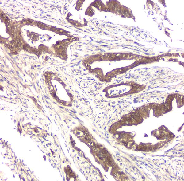
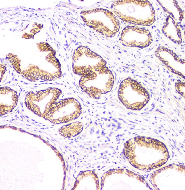
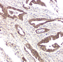
ARG43090 anti-BAG6 / BAT3 antibody IHC-P image
Immunohistochemistry: Paraffin-embedded Human prostatic cancer tissue. Antigen Retrieval: Heat mediation was performed in Citrate buffer (pH 6.0) for 20 min. The tissue section was blocked with 10% goat serum. The tissue section was then stained with ARG43090 anti-BAG6 / BAT3 antibody at 1 µg/ml dilution, overnight at 4°C.
-
ARG43090 anti-BAG6 / BAT3 antibody IHC-P image
Immunohistochemistry: Paraffin-embedded Human thyroid cancer tissue. Antigen Retrieval: Heat mediation was performed in Citrate buffer (pH 6.0) for 20 min. The tissue section was blocked with 10% goat serum. The tissue section was then stained with ARG43090 anti-BAG6 / BAT3 antibody at 1 µg/ml dilution, overnight at 4°C.
-

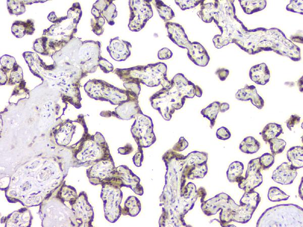
ARG43090 anti-BAG6 / BAT3 antibody IHC-P image
Immunohistochemistry: Paraffin-embedded Human placenta tissue. Antigen Retrieval: Heat mediation was performed in Citrate buffer (pH 6.0) for 20 min. The tissue section was blocked with 10% goat serum. The tissue section was then stained with ARG43090 anti-BAG6 / BAT3 antibody at 1 µg/ml dilution, overnight at 4°C.
-
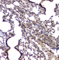
ARG43090 anti-BAG6 / BAT3 antibody IHC-P image
Immunohistochemistry: Paraffin-embedded Mouse lung tissue. Antigen Retrieval: Heat mediation was performed in Citrate buffer (pH 6.0) for 20 min. The tissue section was blocked with 10% goat serum. The tissue section was then stained with ARG43090 anti-BAG6 / BAT3 antibody at 1 µg/ml dilution, overnight at 4°C.
-
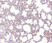
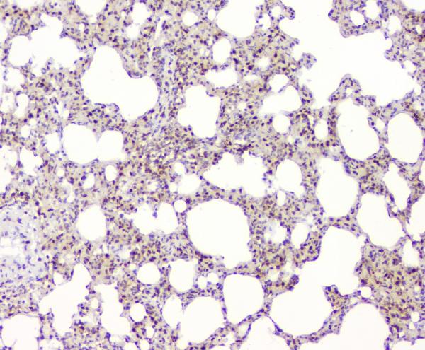
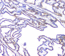
ARG43090 anti-BAG6 / BAT3 antibody IHC-P image
Immunohistochemistry: Paraffin-embedded Rat lung tissue. Antigen Retrieval: Heat mediation was performed in Citrate buffer (pH 6.0) for 20 min. The tissue section was blocked with 10% goat serum. The tissue section was then stained with ARG43090 anti-BAG6 / BAT3 antibody at 1 µg/ml dilution, overnight at 4°C.
-
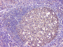
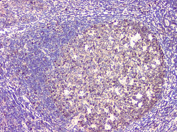
ARG43090 anti-BAG6 / BAT3 antibody IHC-P image
Immunohistochemistry: Paraffin-embedded Human tonsil tissue. Antigen Retrieval: Heat mediation was performed in Citrate buffer (pH 6.0) for 20 min. The tissue section was blocked with 10% goat serum. The tissue section was then stained with ARG43090 anti-BAG6 / BAT3 antibody at 1 µg/ml dilution, overnight at 4°C.
-
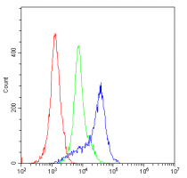
ARG43090 anti-BAG6 / BAT3 antibody FACS image
Flow Cytometry: H-PBMC cells were blocked with 10% normal goat serum and then stained with ARG43090 anti-BAG6 / BAT3 antibody (blue) at 1 µg/10^6 cells for 30 min at 20°C, followed by incubation with DyLight®488 labelled secondary antibody. Isotype control antibody (green) was rabbit IgG (1 µg/10^6 cells) used under the same conditions. Unlabelled sample (red) was also used as a control.