ARG10700
anti-Aurora B antibody [3F11]
anti-Aurora B antibody [3F11] for ICC/IF,IHC-Frozen sections,Western blot and Human,Mouse,Rat,Cow,Horse
Overview
| Product Description | Mouse Monoclonal antibody [3F11] recognizes Aurora B |
|---|---|
| Tested Reactivity | Hu, Ms, Rat, Cow, Hrs |
| Tested Application | ICC/IF, IHC-Fr, WB |
| Specificity | This antibody was made against Aurora B protein and was shown not to bind both Aurora A and C. |
| Host | Mouse |
| Clonality | Monoclonal |
| Clone | 3F11 |
| Isotype | IgG2a |
| Target Name | Aurora B |
| Antigen Species | Human |
| Immunogen | Full length recombinant Human Aurora B protein expressed in and purified from E. coli. |
| Conjugation | Un-conjugated |
| Alternate Names | STK12; PPP1R48; Aurora/IPL1-related kinase 2; Serine/threonine-protein kinase 5; STK-1; IPL1; ARK-2; Aurora kinase B; Aurora-related kinase 2; AIM-1; AIK2; Serine/threonine-protein kinase 12; AIM1; STK5; ARK2; aurkb-sv1; aurkb-sv2; Serine/threonine-protein kinase aurora-B; AurB; Aurora- and IPL1-like midbody-associated protein 1; EC 2.7.11.1; Aurora 1 |
Application Instructions
| Application Suggestion |
|
||||||||
|---|---|---|---|---|---|---|---|---|---|
| Application Note | * The dilutions indicate recommended starting dilutions and the optimal dilutions or concentrations should be determined by the scientist. |
Properties
| Form | Liquid |
|---|---|
| Purification | Affinity purification. |
| Buffer | PBS and 50% Glycerol. |
| Stabilizer | 50% Glycerol |
| Concentration | 1 mg/ml |
| Storage Instruction | For continuous use, store undiluted antibody at 2-8°C for up to a week. For long-term storage, aliquot and store at -20°C. Storage in frost free freezers is not recommended. Avoid repeated freeze/thaw cycles. Suggest spin the vial prior to opening. The antibody solution should be gently mixed before use. |
| Note | For laboratory research only, not for drug, diagnostic or other use. |
Bioinformation
| Database Links | |
|---|---|
| Gene Symbol | AURKB |
| Gene Full Name | aurora kinase B |
| Background | This gene encodes a member of the aurora kinase subfamily of serine/threonine kinases. The genes encoding the other two members of this subfamily are located on chromosomes 19 and 20. These kinases participate in the regulation of alignment and segregation of chromosomes during mitosis and meiosis through association with microtubules. A pseudogene of this gene is located on chromosome 8. Alternatively spliced transcript variants have been found for this gene. [provided by RefSeq, Sep 2015] |
| Function | Serine/threonine-protein kinase component of the chromosomal passenger complex (CPC), a complex that acts as a key regulator of mitosis. The CPC complex has essential functions at the centromere in ensuring correct chromosome alignment and segregation and is required for chromatin-induced microtubule stabilization and spindle assembly. Involved in the bipolar attachment of spindle microtubules to kinetochores and is a key regulator for the onset of cytokinesis during mitosis. Required for central/midzone spindle assembly and cleavage furrow formation. Key component of the cytokinesis checkpoint, a process required to delay abscission to prevent both premature resolution of intercellular chromosome bridges and accumulation of DNA damage: phosphorylates CHMP4C, leading to retain abscission-competent VPS4 (VPS4A and/or VPS4B) at the midbody ring until abscission checkpoint signaling is terminated at late cytokinesis. AURKB phosphorylates the CPC complex subunits BIRC5/survivin, CDCA8/borealin and INCENP. Phosphorylation of INCENP leads to increased AURKB activity. Other known AURKB substrates involved in centromeric functions and mitosis are CENPA, DES/desmin, GPAF, KIF2C, NSUN2, RACGAP1, SEPT1, VIM/vimentin, GSG2/Haspin, and histone H3. A positive feedback loop involving GSG2 and AURKB contributes to localization of CPC to centromeres. Phosphorylation of VIM controls vimentin filament segregation in cytokinetic process, whereas histone H3 is phosphorylated at 'Ser-10' and 'Ser-28' during mitosis (H3S10ph and H3S28ph, respectively). A positive feedback between GSG2 and AURKB contributes to CPC localization. AURKB is also required for kinetochore localization of BUB1 and SGOL1. Phosphorylation of p53/TP53 negatively regulates its transcriptional activity. Key regulator of active promoters in resting B- and T-lymphocytes: acts by mediating phosphorylation of H3S28ph at active promoters in resting B-cells, inhibiting RNF2/RING1B-mediated ubiquitination of histone H2A and enhancing binding and activity of the USP16 deubiquitinase at transcribed genes. [UniProt] |
| Calculated MW | 39 kDa |
| PTM | The phosphorylation of Thr-232 requires the binding to INCENP and occurs by means of an autophosphorylation mechanism. Thr-232 phosphorylation is indispensable for the AURKB kinase activity. Ubiquitinated by different BCR (BTB-CUL3-RBX1) E3 ubiquitin ligase complexes. Ubiquitinated by the BCR(KLHL9-KLHL13) E3 ubiquitin ligase complex, ubiquitination leads to removal from mitotic chromosomes and is required for cytokinesis. During anaphase, the BCR(KLHL21) E3 ubiquitin ligase complex recruits the CPC complex from chromosomes to the spindle midzone and mediates the ubiquitination of AURKB. Ubiquitination of AURKB by BCR(KLHL21) E3 ubiquitin ligase complex may not lead to its degradation by the proteasome. |
Images (4) Click the Picture to Zoom In
-
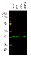
ARG10700 anti-Aurora B antibody [3F11] WB image
Western blot: Cells were treated with 100 ng/ml of nocodazol, a microtubule depolymerizing agent which induces cells to halt at G2/M phase. HeLa, Dog A72, Horse NBL6 and Mouse KR158 cell lysates stained with ARG10700 anti-Aurora B antibody [3F11].
-
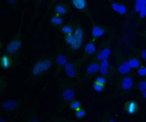
ARG10700 anti-Aurora B antibody [3F11] ICC/IF image
Immunocytochemistry: HeLa cell cultures were stained with ARG10700 anti-Aurora B antibody [3F11] (green).
-
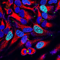
ARG10700 anti-Aurora B antibody [3F11] ICC/IF image
Immunofluorescence: HeLa cells stained with ARG10700 anti-Aurora B antibody [3F11] (green) at 1:1000 dilution and costained with ARG52468 anti-Vimentin antibody (red) at 1:1000 dilution. DAPI (blue) for nuclear staining.
Clone 3F11 reveals Aurora B localized in midbodies, midzones of dividing cells and also in the nuclei or some cells.
-
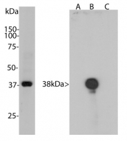
ARG10700 anti-Aurora B antibody [3F11] WB image
Western blot: Left: HeLa cells treated with 100 ng/ml nocodazole for 18 hours was stained with ARG10700 anti-Aurora B antibody [3F11]. Nocodazole is a microtubule polymerization inhibitor which induces cells to halt at the G2/M phase and also induces Aurora B expression. Right: Recombinant Human Aurora A, B and C were stained with ARG10700 anti-Aurora B antibody [3F11]. This antibody binds specifically to Aurora B.


