ARG11120
anti-Aurora A antibody [1A11]
anti-Aurora A antibody [1A11] for ICC/IF,IHC-Frozen sections,Western blot and Human,Mouse
Overview
| Product Description | Mouse Monoclonal antibody [1A11] recognizes Aurora A |
|---|---|
| Tested Reactivity | Hu, Ms |
| Species Does Not React With | Dog, Hrs |
| Tested Application | ICC/IF, IHC-Fr, WB |
| Specificity | This antibody was made against recombinant human aurora A, and was shown to be non-reactive with aurora B or C. |
| Host | Mouse |
| Clonality | Monoclonal |
| Clone | 1A11 |
| Isotype | IgG1 |
| Target Name | Aurora A |
| Antigen Species | Human |
| Immunogen | Recombinant full-length Human Aurora A. |
| Conjugation | Un-conjugated |
| Alternate Names | ARK-1; AIK; BTAK; Serine/threonine-protein kinase 6; Breast tumor-amplified kinase; Serine/threonine-protein kinase aurora-A; STK15; Serine/threonine-protein kinase 15; AURORA2; Aurora-related kinase 1; hARK1; AURA; STK6; STK7; Aurora kinase A; EC 2.7.11.1; Aurora/IPL1-related kinase 1; Aurora 2; ARK1; PPP1R47 |
Application Instructions
| Application Suggestion |
|
||||||||
|---|---|---|---|---|---|---|---|---|---|
| Application Note | * The dilutions indicate recommended starting dilutions and the optimal dilutions or concentrations should be determined by the scientist. | ||||||||
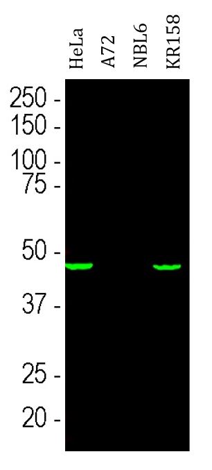
| Observed Size | ~ 46 kDa |
Properties
| Form | Liquid |
|---|---|
| Buffer | Concentrated hybridoma cell culture media and 5 mM Sodium azide. |
| Preservative | 5 mM Sodium azide |
| Storage Instruction | For continuous use, store undiluted antibody at 2-8°C for up to a week. For long-term storage, aliquot and store at -20°C or below. Storage in frost free freezers is not recommended. Avoid repeated freeze/thaw cycles. Suggest spin the vial prior to opening. The antibody solution should be gently mixed before use. |
| Note | For laboratory research only, not for drug, diagnostic or other use. |
Bioinformation
| Database Links | |
|---|---|
| Gene Symbol | AURKA |
| Gene Full Name | aurora kinase A |
| Background | The protein encoded by this gene is a cell cycle-regulated kinase that appears to be involved in microtubule formation and/or stabilization at the spindle pole during chromosome segregation. The encoded protein is found at the centrosome in interphase cells and at the spindle poles in mitosis. This gene may play a role in tumor development and progression. A processed pseudogene of this gene has been found on chromosome 1, and an unprocessed pseudogene has been found on chromosome 10. Multiple transcript variants encoding the same protein have been found for this gene. [provided by RefSeq, Jul 2008] |
| Function | Mitotic serine/threonine kinase that contributes to the regulation of cell cycle progression (PubMed:26246606). Associates with the centrosome and the spindle microtubules during mitosis and plays a critical role in various mitotic events including the establishment of mitotic spindle, centrosome duplication, centrosome separation as well as maturation, chromosomal alignment, spindle assembly checkpoint, and cytokinesis (PubMed:26246606). Required for normal spindle positioning during mitosis and for the localization of NUMA1 and DCTN1 to the cell cortex during metaphase (PubMed:27335426). Required for initial activation of CDK1 at centrosomes. Phosphorylates numerous target proteins, including ARHGEF2, BORA, BRCA1, CDC25B, DLGP5, HDAC6, KIF2A, LATS2, NDEL1, PARD3, PPP1R2, PLK1, RASSF1, TACC3, p53/TP53 and TPX2. Regulates KIF2A tubulin depolymerase activity. Required for normal axon formation. Plays a role in microtubule remodeling during neurite extension. Important for microtubule formation and/or stabilization. Also acts as a key regulatory component of the p53/TP53 pathway, and particularly the checkpoint-response pathways critical for oncogenic transformation of cells, by phosphorylating and stabilizing p53/TP53. Phosphorylates its own inhibitors, the protein phosphatase type 1 (PP1) isoforms, to inhibit their activity. Necessary for proper cilia disassembly prior to mitosis. Regulates protein levels of the anti-apoptosis protein BIRC5 by suppressing the expression of the SCF(FBXL7) E3 ubiquitin-protein ligase substrate adapter FBXL7 through the phosphorylation of the transcription factor FOXP1 (PubMed:28218735). [UniProt] |
| Cellular Localization | Cytoplasm, cytoskeleton, microtubule organizing center, centrosome. Cytoplasm, cytoskeleton, spindle pole. Cytoplasm, cytoskeleton, cilium basal body. Cytoplasm, cytoskeleton, microtubule organizing center, centrosome, centriole. Note=Detected at the neurite hillock in developing neurons. Localizes at the centrosome in mitotic cells from early prophase until telophase, but also localizes to the spindle pole MTs from prophase to anaphase. [UniProt] |
| Calculated MW | 46 kDa |
| PTM | Activated by phosphorylation at Thr-288; this brings about a change in the conformation of the activation segment. Phosphorylation at Thr-288 varies during the cell cycle and is highest during M phase. Autophosphorylated at Thr-288 upon TPX2 binding. Thr-288 can be phosphorylated by several kinases, including PAK and PKA. Protein phosphatase type 1 (PP1) binds AURKA and inhibits its activity by dephosphorylating Thr-288 during mitosis. Phosphorylation at Ser-342 decreases the kinase activity. PPP2CA controls degradation by dephosphorylating Ser-51 at the end of mitosis. Ubiquitinated by the E3 ubiquitin-protein ligase complex SCF(FBXL7) during mitosis, leading to its degradation by the proteasome. Ubiquitinated by CHFR, leading to its degradation by the proteasome (By similarity). Ubiquitinated by the anaphase-promoting complex (APC), leading to its degradation by the proteasome. [UniProt] |
Images (2) Click the Picture to Zoom In
-
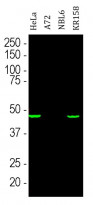
ARG11120 anti-Aurora A antibody [1A11] WB image
Western blot: HeLa, Dog A72 cells, Horse NBL6 cells and Murine KR158 cells. Cells were treated with 100 ng/ml of Nocodazol for 6 hours. Cell lysates were stained with ARG11120 anti-Aurora A antibody [1A11].
-

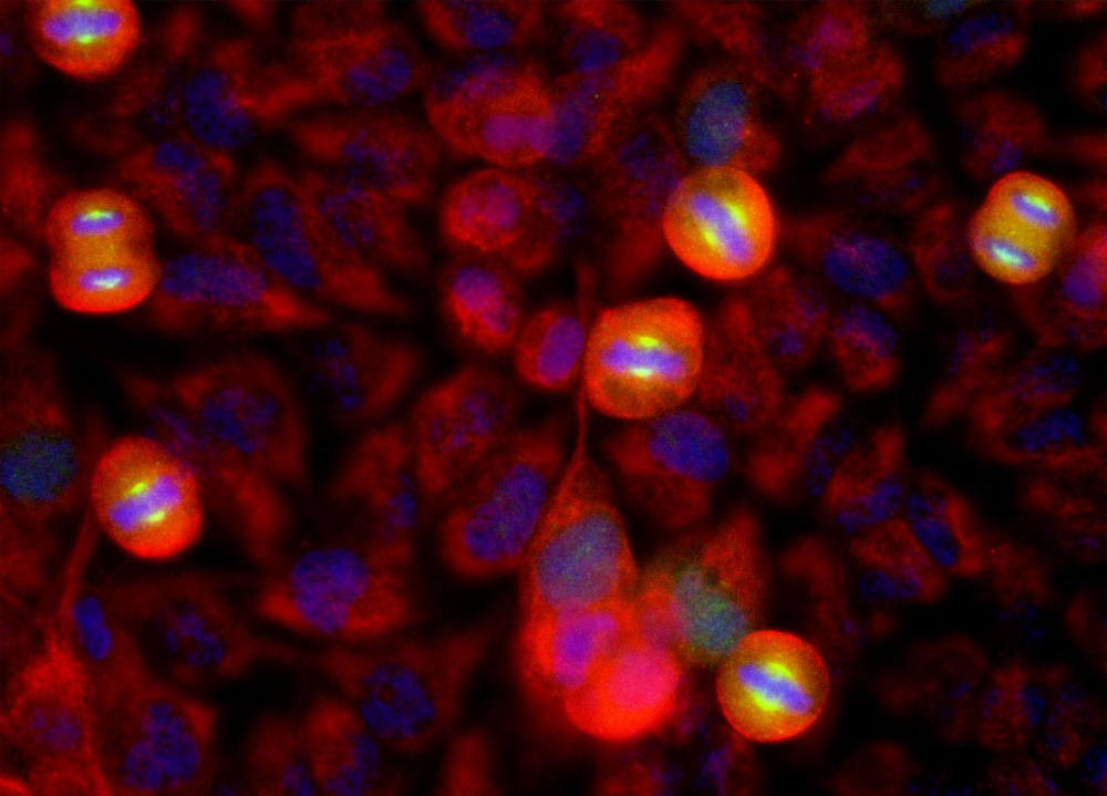
ARG11120 anti-Aurora A antibody [1A11] ICC/IF image
Immunofluorescence: HeLa cells stained with ARG11120 anti-Aurora A antibody [1A11] (green), and co-stained with anti-Vimentin antibody (red). DAPI (blue) for nuclear staining.