ARG10696
anti-Arrestin 1 / S-Arrestin antibody [S128]
anti-Arrestin 1 / S-Arrestin antibody [S128] for ICC/IF,IHC-Frozen sections,Western blot and Bovine,Pig
Overview
| Product Description | Mouse Monoclonal antibody [S128] recognizes Arrestin 1 / S-Arrestin |
|---|---|
| Tested Reactivity | Bov, Pig |
| Tested Application | ICC/IF, IHC-Fr, WB |
| Host | Mouse |
| Clonality | Monoclonal |
| Clone | S128 |
| Isotype | IgG1 |
| Target Name | Arrestin 1 / S-Arrestin |
| Antigen Species | Bovine |
| Immunogen | Recombinant Bovine Arrestin-1 with the first 20 aa of the C-terminus truncated. |
| Conjugation | Un-conjugated |
| Alternate Names | RP47; 48 kDa protein; S-arrestin; Rod photoreceptor arrestin; S-AG; Retinal S-antigen |
Application Instructions
| Application Suggestion |
|
||||||||
|---|---|---|---|---|---|---|---|---|---|
| Application Note | * The dilutions indicate recommended starting dilutions and the optimal dilutions or concentrations should be determined by the scientist. |
Properties
| Form | Liquid |
|---|---|
| Purification | Affinity purification. |
| Buffer | PBS and 50% Glycerol. |
| Stabilizer | 50% Glycerol |
| Concentration | 1 mg/ml |
| Storage Instruction | For continuous use, store undiluted antibody at 2-8°C for up to a week. For long-term storage, aliquot and store at -20°C. Storage in frost free freezers is not recommended. Avoid repeated freeze/thaw cycles. Suggest spin the vial prior to opening. The antibody solution should be gently mixed before use. |
| Note | For laboratory research only, not for drug, diagnostic or other use. |
Bioinformation
| Database Links | |
|---|---|
| Gene Symbol | SAG |
| Gene Full Name | S-antigen; retina and pineal gland (arrestin) |
| Background | Members of arrestin/beta-arrestin protein family are thought to participate in agonist-mediated desensitization of G-protein-coupled receptors and cause specific dampening of cellular responses to stimuli such as hormones, neurotransmitters, or sensory signals. S-arrestin, also known as S-antigen, is a major soluble photoreceptor protein that is involved in desensitization of the photoactivated transduction cascade. It is expressed in the retina and the pineal gland and inhibits coupling of rhodopsin to transducin in vitro. Additionally, S-arrestin is highly antigenic, and is capable of inducing experimental autoimmune uveoretinitis. Mutations in this gene have been associated with Oguchi disease, a rare autosomal recessive form of night blindness. [provided by RefSeq, Jul 2008] |
| Function | Arrestin is one of the major proteins of the ros (retinal rod outer segments); it binds to photoactivated-phosphorylated rhodopsin, thereby apparently preventing the transducin-mediated activation of phosphodiesterase. [UniProt] |
| Calculated MW | 45 kDa |
Images (2) Click the Picture to Zoom In
-
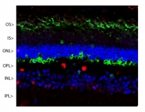
ARG10696 anti-Arrestin 1 / S-Arrestin antibody [S128] IHC-Fr image
Immunohistochemistry: Confocal image of a pig retina frozen section stained with ARG10696 anti-Arrestin 1 / S-Arrestin antibody [S128] (green). Visual arrestin is most abundant in the outer segments (OS) and inner surface of the outer nuclear layer (ONL), and can be used to identify components of rod photoreceptor cells. (Cone photoreceptors have a different arrestin isotype). Other retinal layers are inner segments (IS), outer plexiform layer (OPL), inner nuclear layer (INL) and inner plexiform layer (IPL). The red stain is Fox2, an RNA binding nuclear protein related to Fox3 / NeuN, which stains nuclei of horizontal neurons and some other neurons in the INL and IPL. Nuclear DNA was revealed with DAPI (blue).
-
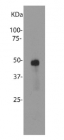
ARG10696 anti-Arrestin 1 / S-Arrestin antibody [S128] WB image
Western blot: Bovine retinal extracts stained with ARG10696 anti-Arrestin 1 / S-Arrestin antibody [S128].


