ARG41560
anti-Aquaporin 1 antibody
anti-Aquaporin 1 antibody for Flow cytometry,IHC-Frozen sections,IHC-Formalin-fixed paraffin-embedded sections,Western blot and Human,Mouse,Rat
Overview
| Product Description | Rabbit Polyclonal antibody recognizes Aquaporin 1 |
|---|---|
| Tested Reactivity | Hu, Ms, Rat |
| Predict Reactivity | Hm |
| Tested Application | FACS, IHC-Fr, IHC-P, WB |
| Host | Rabbit |
| Clonality | Polyclonal |
| Isotype | IgG |
| Target Name | Aquaporin 1 |
| Antigen Species | Human |
| Immunogen | Synthetic peptide corresponding to aa. 251-269 of Human Aquaporin 1. (EEYDLDADDINSRVEMKPK) |
| Conjugation | Un-conjugated |
| Alternate Names | Aquaporin-1; CO; AQP-CHIP; AQP-1; Aquaporin-CHIP; Water channel protein for red blood cells and kidney proximal tubule; CHIP28; Urine water channel |
Application Instructions
| Application Suggestion |
|
||||||||||
|---|---|---|---|---|---|---|---|---|---|---|---|
| Application Note | IHC-P: Antigen Retrieval: Heat mediation was performed in Citrate buffer (pH 6.0) for 20 min, or performed in EDTA buffer (pH 8.0). * The dilutions indicate recommended starting dilutions and the optimal dilutions or concentrations should be determined by the scientist. |
||||||||||
| Observed Size | ~ 30 kDa |
Properties
| Form | Liquid |
|---|---|
| Purification | Affinity purification with immunogen. |
| Buffer | 0.2% Na2HPO4, 0.9% NaCl, 0.05% Thimerosal, 0.05% Sodium azide and 5% BSA. |
| Preservative | 0.05% Thimerosal and 0.05% Sodium azide |
| Stabilizer | 5% BSA |
| Concentration | 0.5 mg/ml |
| Storage Instruction | For continuous use, store undiluted antibody at 2-8°C for up to a week. For long-term storage, aliquot and store at -20°C or below. Storage in frost free freezers is not recommended. Avoid repeated freeze/thaw cycles. Suggest spin the vial prior to opening. The antibody solution should be gently mixed before use. |
| Note | For laboratory research only, not for drug, diagnostic or other use. |
Bioinformation
| Database Links | |
|---|---|
| Gene Symbol | AQP1 |
| Gene Full Name | aquaporin 1 (Colton blood group) |
| Background | Aquaporins are a family of small integral membrane proteins related to the major intrinsic protein (MIP or AQP0). This gene encodes an aquaporin which functions as a molecular water channel protein. It is a homotetramer with 6 bilayer spanning domains and N-glycosylation sites. The protein physically resembles channel proteins and is abundant in erythrocytes and renal tubes. The gene encoding this aquaporin is a possible candidate for disorders involving imbalance in ocular fluid movement. Several transcript variants encoding different isoforms have been found for this gene. [provided by RefSeq, Jun 2010] |
| Function | Forms a water-specific channel that provides the plasma membranes of red cells and kidney proximal tubules with high permeability to water, thereby permitting water to move in the direction of an osmotic gradient. [UniProt] |
| Cellular Localization | Cell membrane; Multi-pass membrane protein. [UniProt] |
| Calculated MW | 29 kDa |
Images (7) Click the Picture to Zoom In
-
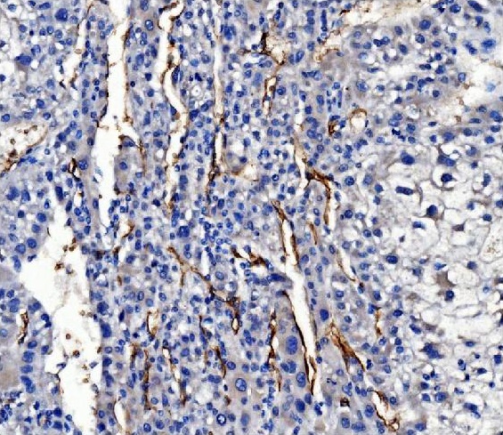
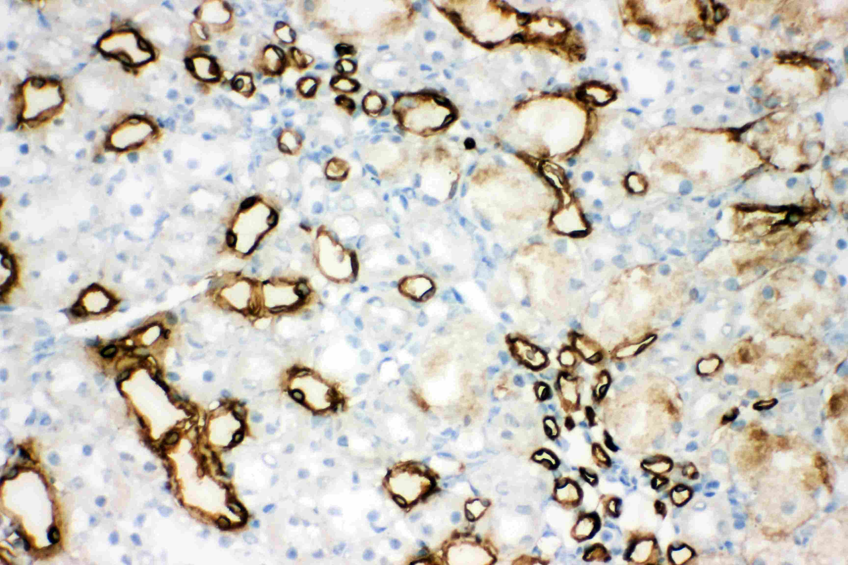
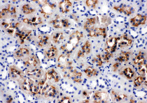
ARG41560 anti-Aquaporin 1 antibody IHC-P image
Immunohistochemistry: Paraffin-embedded Human renal cancer tissue. Antigen Retrieval: Heat mediation was performed in Citrate buffer (pH 6.0, epitope retrieval solution) for 20 min. The tissue section was blocked with 10% goat serum. The tissue section was then stained with ARG41560 anti-Aquaporin 1 antibody at 1 µg/ml dilution, overnight at 4°C.
-
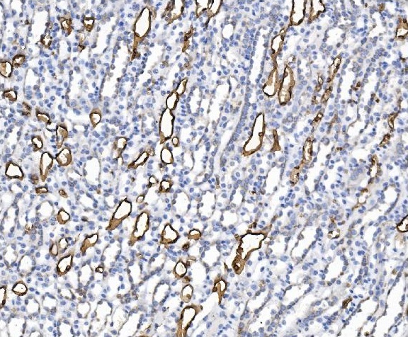
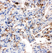
ARG41560 anti-Aquaporin 1 antibody IHC-P image
Immunohistochemistry: Paraffin-embedded Human liver cancer tissue. Antigen Retrieval: Heat mediation was performed in EDTA buffer (pH 8.0). The tissue section was blocked with 10% goat serum. The tissue section was then stained with ARG41560 anti-Aquaporin 1 antibody at 1 µg/ml dilution, overnight at 4°C.
-
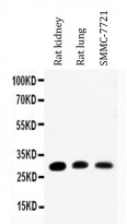
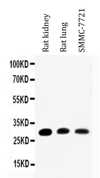
ARG41560 anti-Aquaporin 1 antibody WB image
Western blot: 50 µg of samples under reducing conditions. Rat kidney, Rat lung and SMMC-7721 cell lysates stained with ARG41560 anti-Aquaporin 1 antibody at 0.5 µg/ml dilution, overnight at 4°C.
-
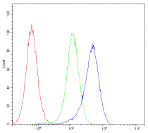
ARG41560 anti-Aquaporin 1 antibody FACS image
Flow Cytometry: U2OS cells were blocked with 10% normal Goat serum and then stained with ARG41560 anti-Aquaporin 1 antibody (blue) at 1 µg/10^6 cells for 30 min at 20°C, followed by incubation with DyLight®488 labelled secondary antibody. Isotype control antibody (green) was Rabbit IgG (1 µg/10^6 cells) used under the same conditions. Unlabelled sample (red) was also used as a control.
-
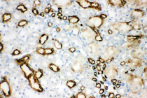
ARG41560 anti-Aquaporin 1 antibody IHC-P image
Immunohistochemistry: Paraffin-embedded Rat kidney tissue. Antigen Retrieval: Heat mediation was performed in Citrate buffer (pH 6.0, epitope retrieval solution) for 20 min. The tissue section was blocked with 10% goat serum. The tissue section was then stained with ARG41560 anti-Aquaporin 1 antibody at 1 µg/ml dilution, overnight at 4°C.
-

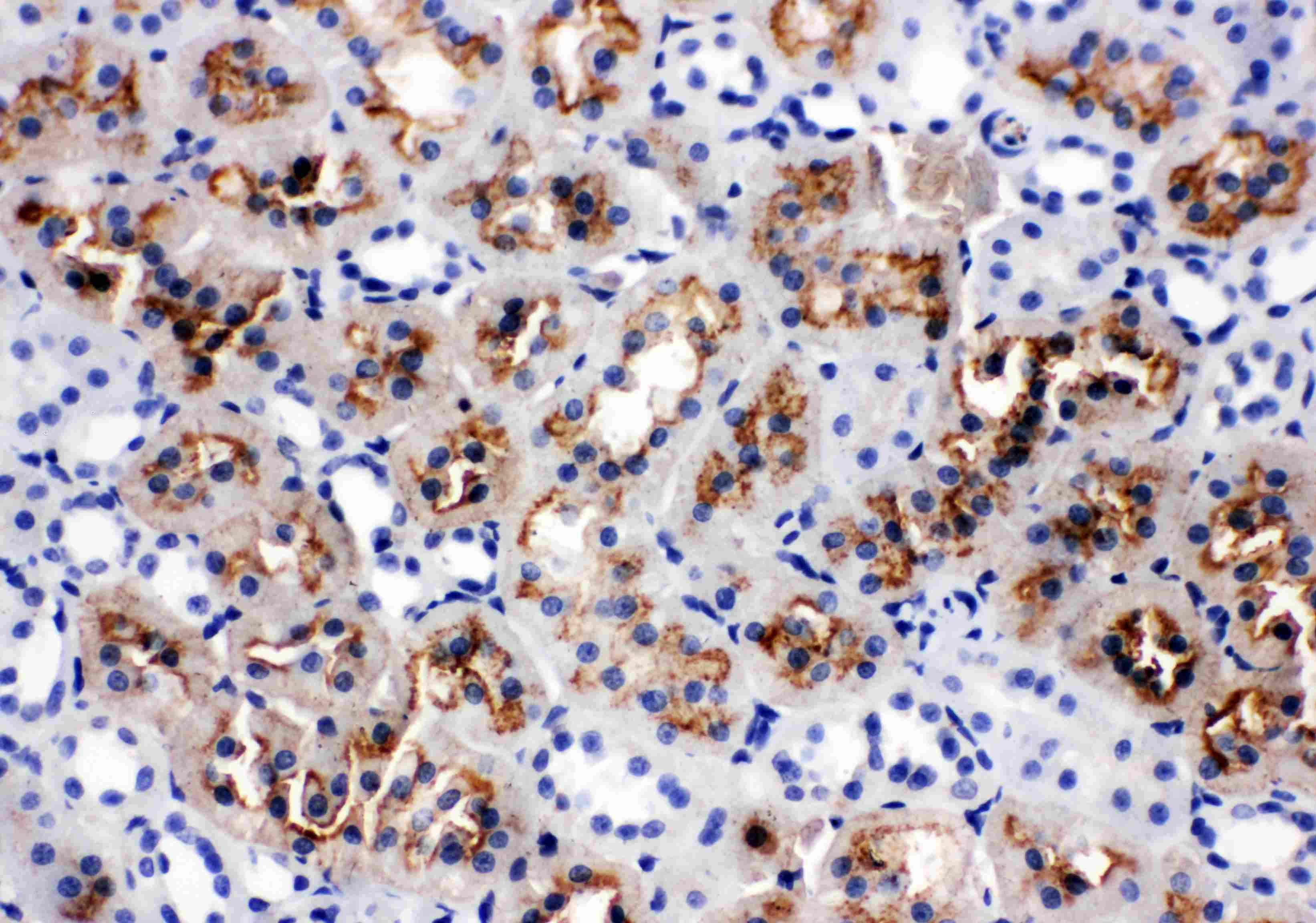
ARG41560 anti-Aquaporin 1 antibody IHC-P image
Immunohistochemistry: Paraffin-embedded Mouse kidney tissue. Antigen Retrieval: Heat mediation was performed in EDTA buffer (pH 8.0). The tissue section was blocked with 10% goat serum. The tissue section was then stained with ARG41560 anti-Aquaporin 1 antibody at 1 µg/ml dilution, overnight at 4°C.
-
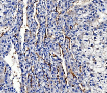
ARG41560 anti-Aquaporin 1 antibody IHC-Fr image
Immunohistochemistry: Frozen section of Rat kidney tissue. The tissue section was blocked with 10% goat serum. The tissue section was then stained with ARG41560 anti-Aquaporin 1 antibody at 1 µg/ml, overnight at 4°C.