ARG66417
anti-ApoJ / Clusterin antibody
anti-ApoJ / Clusterin antibody for ICC/IF,IHC-Formalin-fixed paraffin-embedded sections and Human
Overview
| Product Description | Rabbit Polyclonal antibody recognizes ApoJ / Clusterin |
|---|---|
| Tested Reactivity | Hu |
| Tested Application | ICC/IF, IHC-P |
| Host | Rabbit |
| Clonality | Polyclonal |
| Isotype | IgG |
| Target Name | ApoJ / Clusterin |
| Antigen Species | Human |
| Immunogen | Synthetic peptide corresponding to aa. 370-450 of Human ApoJ / Clusterin. |
| Conjugation | Un-conjugated |
| Alternate Names | Clusterin; Apolipoprotein J; SGP-2; SP-40; APOJ; ApoJbeta; CLU2; CLU1; NA1/NA2; TRPM2; CLI; SGP2; Apo-J; TRPM-2; ApoJalpha; Aging-associated gene 4 protein; APO-J; Complement cytolysis inhibitor a chain; Ku70-binding protein 1; Complement cytolysis inhibitor b chain; Testosterone-repressed prostate message 2; Complement-associated protein SP-40,40; KUB1; AAG4; Complement cytolysis inhibitor |
Application Instructions
| Application Suggestion |
|
||||||
|---|---|---|---|---|---|---|---|
| Application Note | IHC-P: Antigen Retrieval: High-pressure and temperature EDTA buffer (pH 8.0). * The dilutions indicate recommended starting dilutions and the optimal dilutions or concentrations should be determined by the scientist. |
Properties
| Form | Liquid |
|---|---|
| Purification | Affinity purification with immunogen. |
| Buffer | PBS, 0.02% Sodium azide, 50% Glycerol and 0.5% BSA. |
| Preservative | 0.02% Sodium azide |
| Stabilizer | 50% Glycerol and 0.5% BSA |
| Concentration | 1 mg/ml |
| Storage Instruction | For continuous use, store undiluted antibody at 2-8°C for up to a week. For long-term storage, aliquot and store at -20°C. Storage in frost free freezers is not recommended. Avoid repeated freeze/thaw cycles. Suggest spin the vial prior to opening. The antibody solution should be gently mixed before use. |
| Note | For laboratory research only, not for drug, diagnostic or other use. |
Bioinformation
| Database Links | |
|---|---|
| Gene Symbol | CLU |
| Gene Full Name | clusterin |
| Background | The protein encoded by this gene is a secreted chaperone that can under some stress conditions also be found in the cell cytosol. It has been suggested to be involved in several basic biological events such as cell death, tumor progression, and neurodegenerative disorders. Alternate splicing results in both coding and non-coding variants.[provided by RefSeq, May 2011] |
| Function | Isoform 1 functions as extracellular chaperone that prevents aggregation of nonnative proteins. Prevents stress-induced aggregation of blood plasma proteins. Inhibits formation of amyloid fibrils by APP, APOC2, B2M, CALCA, CSN3, SNCA and aggregation-prone LYZ variants (in vitro). Does not require ATP. Maintains partially unfolded proteins in a state appropriate for subsequent refolding by other chaperones, such as HSPA8/HSC70. Does not refold proteins by itself. Binding to cell surface receptors triggers internalization of the chaperone-client complex and subsequent lysosomal or proteasomal degradation. Secreted isoform 1 protects cells against apoptosis and against cytolysis by complement. Intracellular isoforms interact with ubiquitin and SCF (SKP1-CUL1-F-box protein) E3 ubiquitin-protein ligase complexes and promote the ubiquitination and subsequent proteasomal degradation of target proteins. Promotes proteasomal degradation of COMMD1 and IKBKB. Modulates NF-kappa-B transcriptional activity. Nuclear isoforms promote apoptosis. Mitochondrial isoforms suppress BAX-dependent release of cytochrome c into the cytoplasm and inhibit apoptosis. Plays a role in the regulation of cell proliferation. [UniProt] |
| Cellular Localization | Isoform 1: Secreted. Nucleus. Cytoplasm. Mitochondrion membrane; Peripheral membrane protein; Cytoplasmic side. Cytoplasm, cytosol. Microsome. Endoplasmic reticulum. Cytoplasmic vesicle, secretory vesicle, chromaffin granule. [UniProt] |
| Calculated MW | 52 kDa |
| PTM | Isoform 1 is proteolytically cleaved on its way through the secretory system, probably within the Golgi lumen. Polyubiquitinated, leading to proteasomal degradation. Heavily N-glycosylated. About 30% of the protein mass is comprised of complex N-linked carbohydrate. [UniProt] |
Images (3) Click the Picture to Zoom In
-
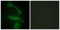
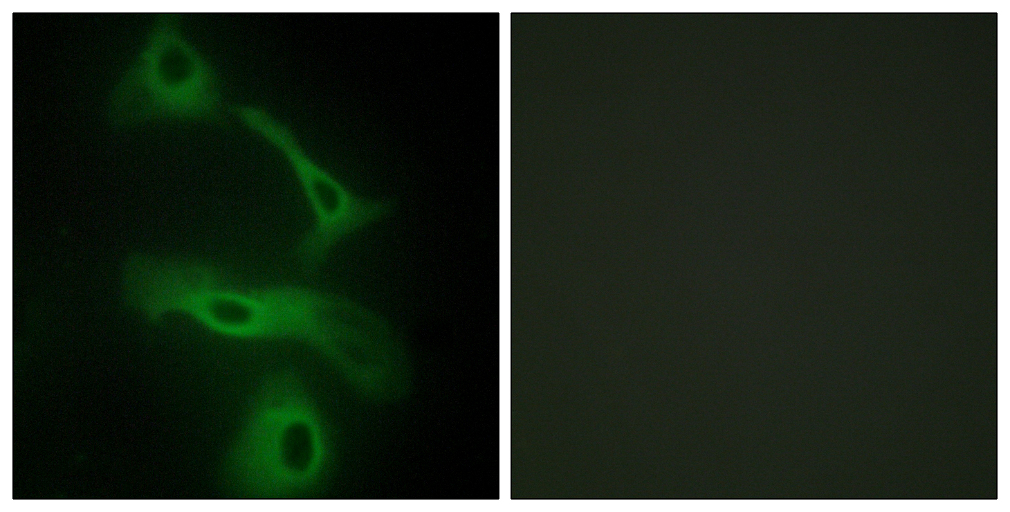
ARG66417 anti-ApoJ / Clusterin antibody ICC/IF image
Immunofluorescence: HeLa cells stained with ARG66417 anti-ApoJ / Clusterin antibody. The picture on the right is blocked with the synthetic peptide.
-
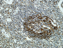
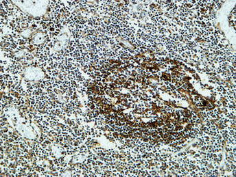
ARG66417 anti-ApoJ / Clusterin antibody IHC-P image
Immunohistochemistry: Paraffin-embedded Human amygdala stained with ARG66417 anti-ApoJ / Clusterin antibody at 1:400 dilution, 4°C and overnight. Antigen Retrieval: High-pressure and temperature EDTA buffer (pH 8.0).
-
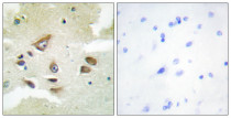
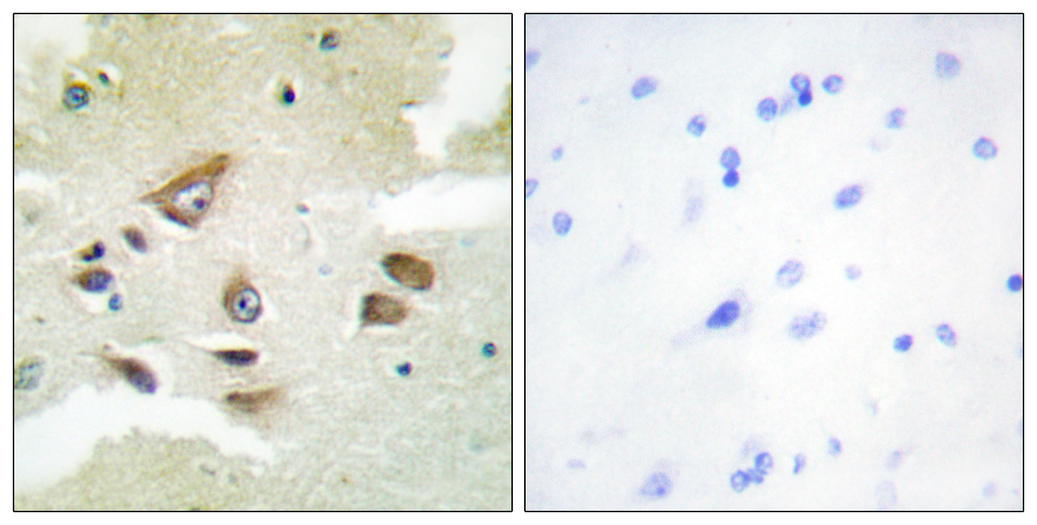
ARG66417 anti-ApoJ / Clusterin antibody IHC-P image
Immunohistochemistry: Paraffin-embedded Human brain stained with ARG66417 anti-ApoJ / Clusterin antibody. The picture on the right is blocked with the synthetic peptide.