ARG10548
anti-Akt 1 antibody [C20-A]
anti-Akt 1 antibody [C20-A] for ICC/IF,Immunoprecipitation,Western blot and Human,Mouse,Rat
Cancer antibody; Cell Death antibody; Gene Regulation antibody; Metabolism antibody; Signaling Transduction antibody; Glucose uptake: Insulin Receptor Dependent Pathway Study antibody
Overview
| Product Description | Rabbit Monoclonal antibody [C20-A] recognizes Akt 1 |
|---|---|
| Tested Reactivity | Hu, Ms, Rat |
| Tested Application | ICC/IF, IP, WB |
| Host | Rabbit |
| Clonality | Monoclonal |
| Clone | C20-A |
| Isotype | IgG |
| Target Name | Akt 1 |
| Antigen Species | Human |
| Immunogen | Synthetic peptide around the C-terminus of Human AKT1 protein |
| Conjugation | Un-conjugated |
| Alternate Names | Protein kinase B alpha; Proto-oncogene c-Akt; RAC; PKB alpha; RAC-ALPHA; CWS6; PRKBA; AKT; PKB; RAC-PK-alpha; PKB-ALPHA; RAC-alpha serine/threonine-protein kinase; EC 2.7.11.1; Protein kinase B |
Application Instructions
| Application Suggestion |
|
||||||||
|---|---|---|---|---|---|---|---|---|---|
| Application Note | * The dilutions indicate recommended starting dilutions and the optimal dilutions or concentrations should be determined by the scientist. |
Properties
| Form | Liquid |
|---|---|
| Buffer | 20 mM Tris-HCl (pH 8.0), 0.05% Sodium azide and 10 mg/ml BSA |
| Preservative | 0.05% Sodium azide |
| Stabilizer | 10 mg/ml BSA |
| Storage Instruction | For continuous use, store undiluted antibody at 2-8°C for up to a week. For long-term storage, aliquot and store at -20°C or below. Storage in frost free freezers is not recommended. Avoid repeated freeze/thaw cycles. Suggest spin the vial prior to opening. The antibody solution should be gently mixed before use. |
| Note | For laboratory research only, not for drug, diagnostic or other use. |
Bioinformation
| Database Links | |
|---|---|
| Gene Symbol | AKT1 |
| Gene Full Name | v-akt murine thymoma viral oncogene homolog 1 |
| Background | The serine-threonine protein kinase encoded by the AKT1 gene is catalytically inactive in serum-starved primary and immortalized fibroblasts. AKT1 and the related AKT2 are activated by platelet-derived growth factor. The activation is rapid and specific, and it is abrogated by mutations in the pleckstrin homology domain of AKT1. It was shown that the activation occurs through phosphatidylinositol 3-kinase. In the developing nervous system AKT is a critical mediator of growth factor-induced neuronal survival. Survival factors can suppress apoptosis in a transcription-independent manner by activating the serine/threonine kinase AKT1, which then phosphorylates and inactivates components of the apoptotic machinery. Mutations in this gene have been associated with the Proteus syndrome. Multiple alternatively spliced transcript variants have been found for this gene. [provided by RefSeq, Jul 2011] |
| Function | Plays a role as a key modulator of the AKT-mTOR signaling pathway controlling the tempo of the process of newborn neurons integration during adult neurogenesis, including correct neuron positioning, dendritic development and synapse formation (By similarity). General protein kinase capable of phosphorylating several known proteins. Phosphorylates TBC1D4. Signals downstream of phosphatidylinositol 3-kinase (PI(3)K) to mediate the effects of various growth factors such as platelet-derived growth factor (PDGF), epidermal growth factor (EGF), insulin and insulin-like growth factor I (IGF-I). Plays a role in glucose transport by mediating insulin-induced translocation of the GLUT4 glucose transporter to the cell surface. Mediates the antiapoptotic effects of IGF-I. Mediates insulin-stimulated protein synthesis by phosphorylating TSC2 at 'Ser-939' and 'Thr-1462', thereby activating mTORC1 signaling and leading to both phosphorylation of 4E-BP1 and in activation of RPS6KB1. Promotes glycogen synthesis by mediating the insulin-induced activation of glycogen synthase. The activated form can suppress FoxO gene transcription and promote cell cycle progression. Essential for the SPATA13-mediated regulation of cell migration and adhesion assembly and disassembly. [UniProt] |
| Research Area | Cancer antibody; Cell Death antibody; Gene Regulation antibody; Metabolism antibody; Signaling Transduction antibody; Glucose uptake: Insulin Receptor Dependent Pathway Study antibody |
| Calculated MW | 56 kDa |
| PTM | O-GlcNAcylation at Thr-305 and Thr-312 inhibits activating phosphorylation at Thr-308 via disrupting the interaction between AKT1 and PDPK1. O-GlcNAcylation at Ser-473 also probably interferes with phosphorylation at this site. Phosphorylation on Thr-308, Ser-473 and Tyr-474 is required for full activity. Activated TNK2 phosphorylates it on Tyr-176 resulting in its binding to the anionic plasma membrane phospholipid PA. This phosphorylated form localizes to the cell membrane, where it is targeted by PDPK1 and PDPK2 for further phosphorylations on Thr-308 and Ser-473 leading to its activation. Ser-473 phosphorylation by mTORC2 favors Thr-308 phosphorylation by PDPK1. Phosphorylated at Thr-308 and Ser-473 by IKBKE and TBK1. Ser-473 phosphorylation is enhanced by interaction with AGAP2 isoform 2 (PIKE-A). Ser-473 phosphorylation is enhanced in focal cortical dysplasias with Taylor-type balloon cells. Ser-473 phosphorylation is enhanced by signaling through activated FLT3. Dephosphorylated at Thr-308 and Ser-473 by PP2A phosphatase. The phosphorylated form of PPP2R5B is required for bridging AKT1 with PP2A phosphatase. Ser-473 is dephosphorylated by CPPED1, leading to termination of signaling. Ubiquitinated via 'Lys-48'-linked polyubiquitination by ZNRF1, leading to its degradation by the proteasome (By similarity). Ubiquitinated; undergoes both 'Lys-48'- and 'Lys-63'-linked polyubiquitination. TRAF6-induced 'Lys-63'-linked AKT1 ubiquitination is critical for phosphorylation and activation. When ubiquitinated, it translocates to the plasma membrane, where it becomes phosphorylated. When fully phosphorylated and translocated into the nucleus, undergoes 'Lys-48'-polyubiquitination catalyzed by TTC3, leading to its degradation by the proteasome. Also ubiquitinated by TRIM13 leading to its proteasomal degradation. Phosphorylated, undergoes 'Lys-48'-linked polyubiquitination preferentially at Lys-284 catalyzed by MUL1, leading to its proteasomal degradation. Acetylated on Lys-14 and Lys-20 by the histone acetyltransferases EP300 and KAT2B. Acetylation results in reduced phosphorylation and inhibition of activity. Deacetylated at Lys-14 and Lys-20 by SIRT1. SIRT1-mediated deacetylation relieves the inhibition. [UniProt] |
Images (3) Click the Picture to Zoom In
-
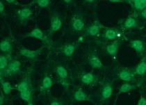
ARG10548 anti-Akt 1 antibody [C20-A] ICC/IF image
Immunocytochemistry: HEK293 cells stained with ARG10548 anti-Akt 1 antibody [C20-A] at 1:300 dilution.
-
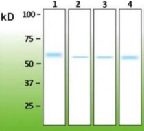
ARG10548 anti-Akt 1 antibody [C20-A] WB image
Western blot: 1) 100 ng of recombinant Human Akt1 (His-tagged), 2) 20 µg, 3) 100 µg, and 4) 200 µg of Mouse brain extracts stained with ARG10548 anti-Akt 1 antibody [C20-A].
-
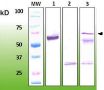
ARG10548 anti-Akt 1 antibody [C20-A] IP image
Immunoprecipitation: 1) 50 ng of recombinant Akt1, 2) 200 µg of Mouse brain extract, immuprecipitation protocol followed without the primary antibody (negative control). 3) Akt1 kinase immunoprecipitation from 200 µg of Mouse brain protein extract with ARG10548 anti-Akt 1 antibody [C20-A].
* Dobransky T et al (2003) Phosphorylation of 69 kDa choline acetyltransferase at threonine-456 in response to short-term exposure to amyloid-ß peptide 1-42. J Biol Chem 278, 5883-5893.


