ARG20523
anti-Acetylated Lysine antibody
anti-Acetylated Lysine antibody for ELISA,ICC/IF,Immunoprecipitation,Western blot and Other
Gene Regulation antibody
Overview
| Product Description | Rabbit Polyclonal antibody recognizes Acetylated Lysine |
|---|---|
| Tested Reactivity | Other |
| Tested Application | ELISA, ICC/IF, IP, WB |
| Specificity | Detects proteins containing acetylated lysine residues. Does not detect non-acetylated lysine residues. |
| Host | Rabbit |
| Clonality | Polyclonal |
| Target Name | Acetylated Lysine |
| Immunogen | Acetylated KLH |
| Conjugation | Un-conjugated |
Application Instructions
| Cross Reactivity Note | Species Independent. | ||||||||||
|---|---|---|---|---|---|---|---|---|---|---|---|
| Application Suggestion |
|
||||||||||
| Application Note | * The dilutions indicate recommended starting dilutions and the optimal dilutions or concentrations should be determined by the scientist. |
Properties
| Form | Liquid |
|---|---|
| Purification | Affinity purification with immunogen. |
| Buffer | PBS, 0.09% Sodium azide and 50% Glycerol |
| Preservative | 0.09% Sodium azide |
| Stabilizer | 50% Glycerol |
| Concentration | 1 mg/ml |
| Storage Instruction | For continuous use, store undiluted antibody at 2-8°C for up to a week. For long-term storage, aliquot and store at -20°C. Storage in frost free freezers is not recommended. Avoid repeated freeze/thaw cycles. Suggest spin the vial prior to opening. The antibody solution should be gently mixed before use. |
| Note | For laboratory research only, not for drug, diagnostic or other use. |
Bioinformation
| Background | Post-translational modifications of proteins play critical roles in the regulation and function of many known biological processes. Proteins can be post-translationally modified in many different ways, and a common post-transcriptional modification of Lysine involves acetylation (1). The conserved amino-terminal domains of the four core histones (H2A, H2B, H3 and H4) contain lysines that are acetylated by histone acetyltransferases (HATs) and deacetylated by histone deacetylases (HDACs) (2). Protein posttranslational reversible lysine Nε-acetylation and deacetylation have been recognized as an emerging intracellular signaling mechanism that plays critical roles in regulating gene transcription, cell-cycle progression, apoptosis, DNA repair, and cytoskeletal organization (3). The regulation of protein acetylation status is impaired in the pathologies of cancer and polyglutamine diseases (4), and HDACs have become promising targets for anti-cancer drugs currently in development (5). 1. Yang XJ. (2005). Oncogene. 24:1653-1662. 2. Hassig, C.A. and Schreiber, S.L. (1997). Curr. Opin. Chem. Biol. 1(3): 300-308. 3. Yang XJ. (2004). Bioessays 26:1076-1087. 4. Hughes, R.E. (2002). Curr. Biol. 12: R141-R143. 5. Vigushin, D.M. and Coombes, R.C. (2004). Curr. Cancer Drug Targets 4: 205-218. 6. Chan, H.M. et al. (2001). Nat. Cell Biol. 3: 667- . 674. 7. Martinez-Balbas, M.A. et al. (2000). EMBO J. 19: 662-671. |
|---|---|
| Research Area | Gene Regulation antibody |
Images (4) Click the Picture to Zoom In
-
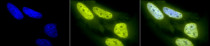
ARG20523 anti-Acetylated Lysine antibody ICC/IF image
Immunofluorescence: Heat Shocked (42°C for 1 hour) HeLa cells. Fixation: 2% Formaldehyde for 20 min at RT. Primary Antibody: ARG20523 anti-Acetylated Lysine antibody at 1:100 for 12 hours at 4°C. Secondary Antibody: R-PE Goat anti-Rabbit (yellow) at 1:200 for 2 hours at RT. Counterstain: DAPI (blue) nuclear stain at 1:40000 for 2 hours at RT. Magnification: 100x. Left: DAPI (blue) nuclear stain. Middle: ARG20523 anti-Acetylated Lysine antibody. Right: Composite.
-
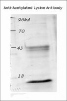
ARG20523 anti-Acetylated Lysine antibody WB image
Western blot: acetylated histone from TSA-treated mouse spleen cells, stained with ARG20523 anti-Acetylated Lysine antibody.
-

ARG20523 anti-Acetylated Lysine antibody ICC/IF image
Immunofluorescence: Heat Shocked (42°C for 1 hour) HeLa cells. Fixation: 2% Formaldehyde for 20 min at RT. Primary Antibody: ARG20523 anti-Acetylated Lysine antibody at 1:100 for 12 hours at 4°C. Secondary Antibody: FITC Goat anti-Rabbit (green) at 1:200 for 2 hours at RT. Counterstain: DAPI (blue) nuclear stain at 1:40000 for 2 hours at RT. Magnification: 20x. Left: DAPI (blue) nuclear stain. Middle: ARG20523 anti-Acetylated Lysine antibody. Right: Composite.
-
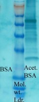
ARG20523 anti-Acetylated Lysine antibody WB image
Western blot: acetylated lysine in BSA (Left) and Acetylated BSA (Right) stained with ARG20523 anti-Acetylated Lysine antibody at 1:1000 dilution.


Specific References
Sirtuin 6 Deacetylates Apoptosis-Associated Speck-Like Protein (ASC) to Inhibit Endothelial Cell Pyroptosis in Atherosclerosis
WB / Human


Interfering TRIB3 protects the blood brain barrier through PI3K/Akt pathway to alleviate cerebral ischemia-reperfusion injury in diabetes mellitus mice
IP / Mouse


