ARG63283
anti-AXIN1 antibody
anti-AXIN1 antibody for IHC-Formalin-fixed paraffin-embedded sections and Human
Cell Biology and Cellular Response antibody; Controls and Markers antibody; Developmental Biology antibody; Signaling Transduction antibody
Overview
| Product Description | Goat Polyclonal antibody recognizes AXIN1 |
|---|---|
| Tested Reactivity | Hu |
| Predict Reactivity | Ms, Rat, Cow, Dog |
| Tested Application | IHC-P |
| Specificity | This antibody is expected to recognise both reported isoforms. |
| Host | Goat |
| Clonality | Polyclonal |
| Isotype | IgG |
| Target Name | AXIN1 |
| Antigen Species | Human |
| Immunogen | C-FEEKIIGKVEKVD |
| Conjugation | Un-conjugated |
| Alternate Names | AXIN; PPP1R49; hAxin; Axin-1; Axis inhibition protein 1 |
Application Instructions
| Application Suggestion |
|
||||
|---|---|---|---|---|---|
| Application Note | IHC-P: Antigen Retrieval: Steam tissue section in Citrate buffer (pH 6.0). * The dilutions indicate recommended starting dilutions and the optimal dilutions or concentrations should be determined by the scientist. |
Properties
| Form | Liquid |
|---|---|
| Purification | Purified from goat serum by ammonium sulphate precipitation followed by antigen affinity chromatography using the immunizing peptide. |
| Buffer | Tris saline (pH 7.3), 0.02% Sodium azide and 0.5% BSA |
| Preservative | 0.02% Sodium azide |
| Stabilizer | 0.5% BSA |
| Concentration | 0.5 mg/ml |
| Storage Instruction | For continuous use, store undiluted antibody at 2-8°C for up to a week. For long-term storage, aliquot and store at -20°C or below. Storage in frost free freezers is not recommended. Avoid repeated freeze/thaw cycles. Suggest spin the vial prior to opening. The antibody solution should be gently mixed before use. |
| Note | For laboratory research only, not for drug, diagnostic or other use. |
Bioinformation
| Database Links | |
|---|---|
| Background | This gene encodes a cytoplasmic protein which contains a regulation of G-protein signaling (RGS) domain and a dishevelled and axin (DIX) domain. The encoded protein interacts with adenomatosis polyposis coli, catenin beta-1, glycogen synthase kinase 3 beta, protein phosphate 2, and itself. This protein functions as a negative regulator of the wingless-type MMTV integration site family, member 1 (WNT) signaling pathway and can induce apoptosis. The crystal structure of a portion of this protein, alone and in a complex with other proteins, has been resolved. Mutations in this gene have been associated with hepatocellular carcinoma, hepatoblastomas, ovarian endometriod adenocarcinomas, and medullablastomas. Two transcript variants encoding distinct isoforms have been identified for this gene. [provided by RefSeq, Dec 2010] |
| Research Area | Cell Biology and Cellular Response antibody; Controls and Markers antibody; Developmental Biology antibody; Signaling Transduction antibody |
| Calculated MW | 96 kDa |
| PTM | Phosphorylation and dephosphorylation of AXIN1 regulates assembly and function of the beta-catenin complex. Phosphorylated by CK1 and GSK3B. Dephosphorylated by PPP1CA and PPP2CA. Phosphorylation by CK1 enhances binding of GSK3B to AXIN1. ADP-ribosylated by tankyrase TNKS and TNKS2. Poly-ADP-ribosylated protein is recognized by RNF146, followed by ubiquitination at 'Lys-48' and subsequent activation of the Wnt signaling pathway. Ubiquitinated by RNF146 when poly-ADP-ribosylated, leading to its degradation and subsequent activation of the Wnt signaling pathway. Sumoylation at Lys-857 and Lys-860 prevents ubiquitination and degradation. Sumoylation is required for AXIN1-mediated JNK activation. Deubiquitinated by USP34, deubiquitinated downstream of beta-catenin stabilization step: deubiquitination is important for nuclear accumulation during Wnt signaling to positively regulate beta-catenin (CTNBB1)-mediated transcription. |
Images (1) Click the Picture to Zoom In
-

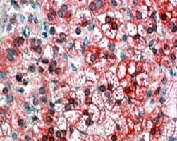
ARG63283 anti-AXIN1 antibody IHC-P image
Immunohistochemistry: paraffin embedded Human Adrenal Cortex. (Steamed antigen retrieval with citrate buffer pH 6) stained with ARG63283 anti-AXIN1 antibody at 3.8 µg/ml dilution followed by AP-staining.