ARG58280
anti-ATP5J antibody
anti-ATP5J antibody for ICC/IF,IHC-Formalin-fixed paraffin-embedded sections,Western blot and Human,Mouse,Rat
Overview
| Product Description | Rabbit Polyclonal antibody recognizes ATP5J |
|---|---|
| Tested Reactivity | Hu, Ms, Rat |
| Predict Reactivity | Mk |
| Tested Application | ICC/IF, IHC-P, WB |
| Host | Rabbit |
| Clonality | Polyclonal |
| Isotype | IgG |
| Target Name | ATP5J |
| Antigen Species | Human |
| Immunogen | KLH-conjugated synthetic peptide corresponding to aa. 28-56 (Center) of Human ATP5J. |
| Conjugation | Un-conjugated |
| Alternate Names | F6; ATP5; ATPM; ATPase subunit F6; CF6; ATP synthase-coupling factor 6, mitochondrial; ATP5A |
Application Instructions
| Application Suggestion |
|
||||||||
|---|---|---|---|---|---|---|---|---|---|
| Application Note | * The dilutions indicate recommended starting dilutions and the optimal dilutions or concentrations should be determined by the scientist. | ||||||||
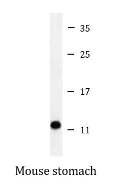
| Positive Control | Mouse stomach |
Properties
| Form | Liquid |
|---|---|
| Purification | Purification with Protein A and immunogen peptide. |
| Buffer | PBS and 0.09% (W/V) Sodium azide. |
| Preservative | 0.09% (W/V) Sodium azide. |
| Storage Instruction | For continuous use, store undiluted antibody at 2-8°C for up to a week. For long-term storage, aliquot and store at -20°C or below. Storage in frost free freezers is not recommended. Avoid repeated freeze/thaw cycles. Suggest spin the vial prior to opening. The antibody solution should be gently mixed before use. |
| Note | For laboratory research only, not for drug, diagnostic or other use. |
Bioinformation
| Database Links | |
|---|---|
| Gene Symbol | ATP5J |
| Gene Full Name | ATP synthase, H+ transporting, mitochondrial Fo complex, subunit F6 |
| Background | Mitochondrial ATP synthase catalyzes ATP synthesis, utilizing an electrochemical gradient of protons across the inner membrane during oxidative phosphorylation. It is composed of two linked multi-subunit complexes: the soluble catalytic core, F1, and the membrane-spanning component, Fo, which comprises the proton channel. The F1 complex consists of 5 different subunits (alpha, beta, gamma, delta, and epsilon) assembled in a ratio of 3 alpha, 3 beta, and a single representative of the other 3. The Fo seems to have nine subunits (a, b, c, d, e, f, g, F6 and 8). This gene encodes the F6 subunit of the Fo complex, required for F1 and Fo interactions. Alternatively spliced transcript variants encoding different isoforms have been identified for this gene. A pseudogene exists on chromosome Yp11.[provided by RefSeq, Jun 2010] |
| Function | Mitochondrial membrane ATP synthase (F(1)F(0) ATP synthase or Complex V) produces ATP from ADP in the presence of a proton gradient across the membrane which is generated by electron transport complexes of the respiratory chain. F-type ATPases consist of two structural domains, F(1) - containing the extramembraneous catalytic core and F(0) - containing the membrane proton channel, linked together by a central stalk and a peripheral stalk. During catalysis, ATP synthesis in the catalytic domain of F(1) is coupled via a rotary mechanism of the central stalk subunits to proton translocation. Part of the complex F(0) domain and the peripheric stalk, which acts as a stator to hold the catalytic alpha(3)beta(3) subcomplex and subunit a/ATP6 static relative to the rotary elements. Also involved in the restoration of oligomycin-sensitive ATPase activity to depleted F1-F0 complexes. [UniProt] |
| Cellular Localization | Mitochondrion. Mitochondrion inner membrane. [UniProt] |
| Calculated MW | 13 kDa |
Images (3) Click the Picture to Zoom In
-
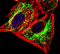
ARG58280 anti-ATP5J antibody ICC/IF image
Immunofluorescence: U251 cells were fixed with 4% PFA (20 min), permeabilized with Triton X-100 (0.1%, 10 min). Cells were stained with ARG58280 anti-ATP5J antibody (green) at 1:25 dilution, 1 hour at 37°C. Cytoplasmic actin was counterstained with Alexa Fluor® 555 (red) conjugated Phalloidin (7 units/ml, 1 h at 37°C). DAPI (blue) for nuclear staining.
-
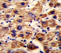
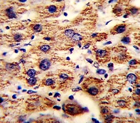
ARG58280 anti-ATP5J antibody IHC-P image
Immunohistochemistry: Formalin-fixed and paraffin-embedded Human liver tissue stained with ARG58280 anti-ATP5J antibody.
-

ARG58280 anti-ATP5J antibody WB image
Western blot: 35 µg of Mouse stomach lysate stained with ARG58280 anti-ATP5J antibody.