ARG58311
anti-ATP5H antibody
anti-ATP5H antibody for ICC/IF,IHC-Formalin-fixed paraffin-embedded sections,Western blot and Human,Mouse,Rat
Overview
| Product Description | Rabbit Polyclonal antibody recognizes ATP5H |
|---|---|
| Tested Reactivity | Hu, Ms, Rat |
| Tested Application | ICC/IF, IHC-P, WB |
| Host | Rabbit |
| Clonality | Polyclonal |
| Isotype | IgG |
| Target Name | ATP5H |
| Antigen Species | Human |
| Immunogen | Human ATP5H recombinant protein (Position: A2-L161). Human ATP5H shares 81% and 78% amino acid (aa) sequence identity with Mouse and Rat ATP5H, respectively. |
| Conjugation | Un-conjugated |
| Alternate Names | ATPQ; ATP synthase subunit d, mitochondrial; ATPase subunit d |
Application Instructions
| Application Suggestion |
|
||||||||
|---|---|---|---|---|---|---|---|---|---|
| Application Note | IHC-P: Antigen Retrieval: By heat mediation. * The dilutions indicate recommended starting dilutions and the optimal dilutions or concentrations should be determined by the scientist. |
Properties
| Form | Liquid |
|---|---|
| Purification | Affinity purification with immunogen. |
| Buffer | 0.9% NaCl, 0.2% Na2HPO4, 0.05% Sodium azide and 5% BSA. |
| Preservative | 0.05% Sodium azide |
| Stabilizer | 5% BSA |
| Concentration | 0.5 mg/ml |
| Storage Instruction | For continuous use, store undiluted antibody at 2-8°C for up to a week. For long-term storage, aliquot and store at -20°C or below. Storage in frost free freezers is not recommended. Avoid repeated freeze/thaw cycles. Suggest spin the vial prior to opening. The antibody solution should be gently mixed before use. |
| Note | For laboratory research only, not for drug, diagnostic or other use. |
Bioinformation
| Database Links | |
|---|---|
| Gene Symbol | ATP5H |
| Gene Full Name | ATP synthase, H+ transporting, mitochondrial Fo complex, subunit d |
| Background | Mitochondrial ATP synthase catalyzes ATP synthesis, utilizing an electrochemical gradient of protons across the inner membrane during oxidative phosphorylation. It is composed of two linked multi-subunit complexes: the soluble catalytic core, F1, and the membrane-spanning component, Fo, which comprises the proton channel. The F1 complex consists of 5 different subunits (alpha, beta, gamma, delta, and epsilon) assembled in a ratio of 3 alpha, 3 beta, and a single representative of the other 3. The Fo seems to have nine subunits (a, b, c, d, e, f, g, F6 and 8). This gene encodes the d subunit of the Fo complex. Alternatively spliced transcript variants encoding different isoforms have been identified for this gene. In addition, three pseudogenes are located on chromosomes 9, 12 and 15. [provided by RefSeq, Jun 2010] |
| Function | Mitochondrial membrane ATP synthase (F(1)F(0) ATP synthase or Complex V) produces ATP from ADP in the presence of a proton gradient across the membrane which is generated by electron transport complexes of the respiratory chain. F-type ATPases consist of two structural domains, F(1) - containing the extramembraneous catalytic core, and F(0) - containing the membrane proton channel, linked together by a central stalk and a peripheral stalk. During catalysis, ATP synthesis in the catalytic domain of F(1) is coupled via a rotary mechanism of the central stalk subunits to proton translocation. Part of the complex F(0) domain and the peripheric stalk, which acts as a stator to hold the catalytic alpha(3)beta(3) subcomplex and subunit a/ATP6 static relative to the rotary elements. [UniProt] |
| Cellular Localization | Mitochondrion. Mitochondrion inner membrane. [UniProt] |
| Calculated MW | 18 kDa |
Images (6) Click the Picture to Zoom In
-
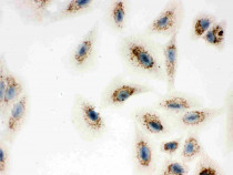
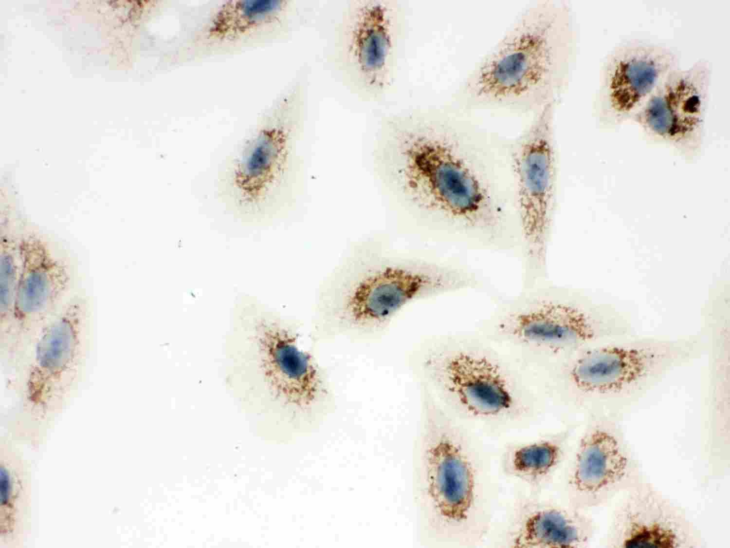
ARG58311 anti-ATP5H antibody ICC image
Immunocytochemistry: A549 cells stained with ARG58311 anti-ATP5H antibody.
-
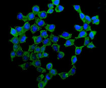
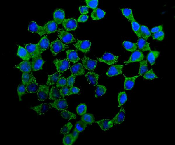
ARG58311 anti-ATP5H antibody ICC/IF image
Immunofluorescence: MCF-7 cells were blocked with 10% goat serum and then stained with ARG58311 anti-ATP5H antibody (green) at 5 µg/ml dilution, overnight at 4°C. DAPI (blue) for nuclear staining.
-
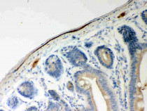
ARG58311 anti-ATP5H antibody IHC-P image
Immunohistochemistry: Paraffin-embedded Mouse intestine stained with ARG58311 anti-ATP5H antibody.
-
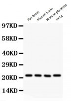
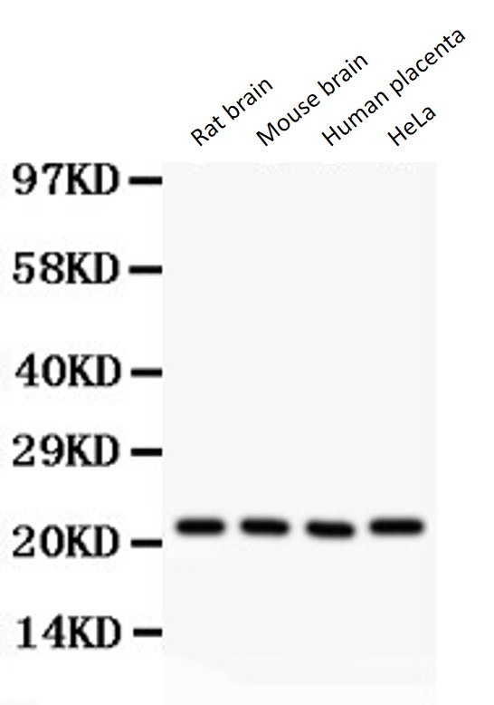
ARG58311 anti-ATP5H antibody WB image
Western blot: 50 µg of Rat brain, 50 µg of Mouse brain, 50 µg of Human placenta and 40 µg of HeLa whole cell lysate stained with ARG58311 anti-ATP5H antibody at 0.5 µg/ml dilution.
-
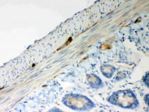
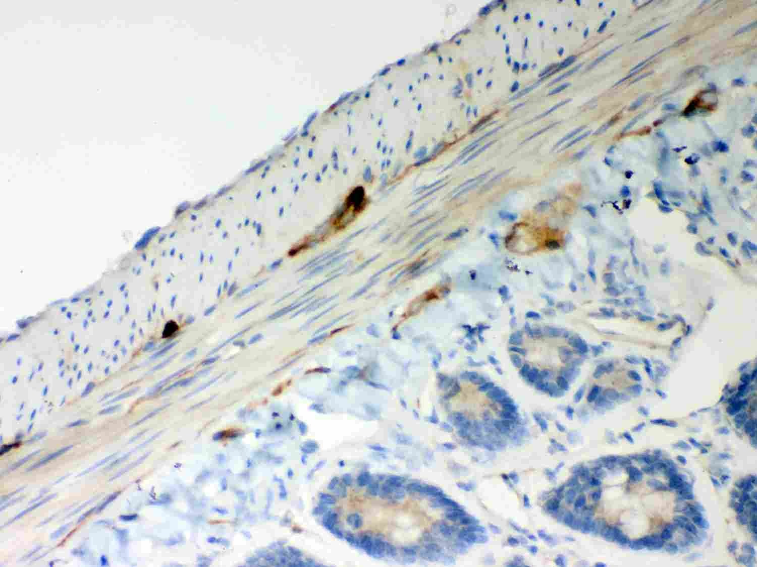
ARG58311 anti-ATP5H antibody IHC-P image
Immunohistochemistry: Paraffin-embedded Rat intestine stained with ARG58311 anti-ATP5H antibody.
-
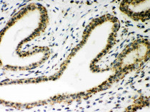
ARG58311 anti-ATP5H antibody IHC-P image
Immunohistochemistry: Paraffin-embedded Human mammary cancer stained with ARG58311 anti-ATP5H antibody.