ARG66994
anti-ATF6 antibody
anti-ATF6 antibody for ICC/IF,IHC-Formalin-fixed paraffin-embedded sections,Western blot and Human,Mouse
Gene Regulation antibody
Overview
| Product Description | Rabbit Polyclonal antibody recognizes ATF6 |
|---|---|
| Tested Reactivity | Hu, Ms |
| Predict Reactivity | Rat |
| Tested Application | ICC/IF, IHC-P, WB |
| Host | Rabbit |
| Clonality | Polyclonal |
| Isotype | IgG |
| Target Name | ATF6 |
| Antigen Species | Human |
| Immunogen | Synthetic peptide around the middle region of Human ATF6. |
| Conjugation | Un-conjugated |
| Alternate Names | ATF6A; cAMP-dependent transcription factor ATF-6 alpha; ATF6-alpha; Activating transcription factor 6 alpha; Cyclic AMP-dependent transcription factor ATF-6 alpha |
Application Instructions
| Application Suggestion |
|
||||||||
|---|---|---|---|---|---|---|---|---|---|
| Application Note | * The dilutions indicate recommended starting dilutions and the optimal dilutions or concentrations should be determined by the scientist. | ||||||||
| Positive Control | MCF7; Human lung; Jurkat | ||||||||
| Observed Size | 72-95 kDa |
Properties
| Form | Liquid |
|---|---|
| Purification | Affinity purified. |
| Buffer | 100 mM Tris Glycine (pH 7.0), 0.025% ProClin 300, 20% Glycerol and 1% BSA. |
| Preservative | 0.025% ProClin 300 |
| Stabilizer | 20% Glycerol and 1% BSA |
| Storage Instruction | For continuous use, store undiluted antibody at 2-8°C for up to a week. For long-term storage, aliquot and store at -20°C or below. Storage in frost free freezers is not recommended. Avoid repeated freeze/thaw cycles. Suggest spin the vial prior to opening. The antibody solution should be gently mixed before use. |
| Note | For laboratory research only, not for drug, diagnostic or other use. |
Bioinformation
| Database Links |
Swiss-port # F6VAN0 Mouse Cyclic AMP-dependent transcription factor ATF-6 alpha Swiss-port # P18850 Human Cyclic AMP-dependent transcription factor ATF-6 alpha |
|---|---|
| Gene Symbol | ATF6 |
| Gene Full Name | activating transcription factor 6 |
| Background | ATF6 Antibody: Disruptions of protein folding and maturation in the endoplasmic reticulum (ER) result in the activation of the unfolded protein response (UPR), an integrated cellular signaling pathway that transmits information from the ER lumen to the cytoplasm and nucleus. Activating transcription factor 6 (ATF6) as well as the ER-transmembrane protein kinases IRE1p and PERK are the major transducers of the UPR. ATF6 is an ER transmembrane protein that is normally bound to the ER chaperone GRP78, but upon ER stress is released from GRP78 and proteolytically cleaved to yield a cytosolic fragment which then migrates to the nucleus, and together with the transcription factor XBP-1, activates transcription of UPR-responsive genes. ATF6 has two isoforms (ATF6α and ATF6β); only ATF6α is recognized by this antibody. |
| Function | Transcription factor that acts during endoplasmic reticulum stress by activating unfolded protein response target genes. Binds DNA on the 5'-CCAC[GA]-3'half of the ER stress response element (ERSE) (5'-CCAAT-N(9)-CCAC[GA]-3') and of ERSE II (5'-ATTGG-N-CCACG-3'). Binding to ERSE requires binding of NF-Y to ERSE. Could also be involved in activation of transcription by the serum response factor. [UniProt] |
| Research Area | Gene Regulation antibody |
| Calculated MW | 75 kDa |
| PTM | During unfolded protein response, a fragment of approximately 50 kDa containing the cytoplasmic transcription factor domain is released by proteolysis. The cleavage seems to be performed sequentially by site-1 and site-2 proteases. N-glycosylated. The glycosylation status may serve as a sensor for ER homeostasis, resulting in ATF6 activation to trigger the unfolded protein response (UPR). Phosphorylated in vitro by MAPK14/P38MAPK. |
Images (3) Click the Picture to Zoom In
-
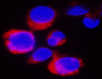
ARG66994 anti-ATF6 antibody IHC-Fr image
Immunofluorescence: Jurkat cells were fixed with 4% paraformaldehyde at RT for 10 min, permeabilized with 0.1% NP-40 at RT for 10 min and then cells were blocked with 5% BSA at RT for 30 min and stained with ARG66994 anti-ATF6 antibody (red) at 1:400 dilution at 4°C for overnight. DAPI (blue) was used for nuclear staining.
-
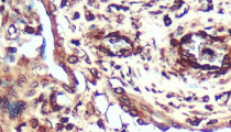
ARG66994 anti-ATF6 antibody IHC-P image
Immunohistochemistry: Paraffin-embedded human lung cancer tissue stained with ARG66994 anti-ATF6 antibody at 1:200 dilution. Antigen Retrieval: Boil tissue section in Citrate buffer (pH 6.0).
-
ARG66994 anti-ATF6 antibody WB image
Western blot: 60 µg of MCF7 cell lysate stained with ARG66994 anti-ATF6 antibody at 1:1000 dilution, overnight at 4°C.


