ARG42610
anti-ATF2 antibody
anti-ATF2 antibody for Flow cytometry,IHC-Frozen sections,IHC-Formalin-fixed paraffin-embedded sections,Western blot and Human,Mouse,Rat
Overview
| Product Description | Rabbit Polyclonal antibody recognizes ATF2 |
|---|---|
| Tested Reactivity | Hu, Ms, Rat |
| Tested Application | FACS, IHC-Fr, IHC-P, WB |
| Host | Rabbit |
| Clonality | Polyclonal |
| Isotype | IgG |
| Target Name | ATF2 |
| Antigen Species | Human |
| Immunogen | Recombinant protein corresponding to E93-E450 of Human ATF2. |
| Conjugation | Un-conjugated |
| Alternate Names | EC 2.3.1.48; Histone acetyltransferase ATF2; Activating transcription factor 2; cAMP-dependent transcription factor ATF-2; Cyclic AMP-dependent transcription factor ATF-2; CREB2; HB16; CREB-2; Cyclic AMP-responsive element-binding protein 2; cAMP-responsive element-binding protein 2; CRE-BP1; cAMP response element-binding protein CRE-BP1; TREB7 |
Application Instructions
| Application Suggestion |
|
||||||||||
|---|---|---|---|---|---|---|---|---|---|---|---|
| Application Note | IHC-P: Antigen Retrieval: Heat mediation was performed in Citrate buffer (pH 6.0) for 20 min. * The dilutions indicate recommended starting dilutions and the optimal dilutions or concentrations should be determined by the scientist. |
Properties
| Form | Liquid |
|---|---|
| Purification | Affinity purification with immunogen. |
| Buffer | 0.2% Na2HPO4, 0.9% NaCl, 0.05% Sodium azide and 5% BSA. |
| Preservative | 0.05% Sodium azide |
| Stabilizer | 5% BSA |
| Concentration | 0.5 mg/ml |
| Storage Instruction | For continuous use, store undiluted antibody at 2-8°C for up to a week. For long-term storage, aliquot and store at -20°C or below. Storage in frost free freezers is not recommended. Avoid repeated freeze/thaw cycles. Suggest spin the vial prior to opening. The antibody solution should be gently mixed before use. |
| Note | For laboratory research only, not for drug, diagnostic or other use. |
Bioinformation
| Database Links | |
|---|---|
| Gene Symbol | ATF2 |
| Gene Full Name | activating transcription factor 2 |
| Background | This gene encodes a transcription factor that is a member of the leucine zipper family of DNA binding proteins. The encoded protein has been identified as a moonlighting protein based on its ability to perform mechanistically distinct functions This protein binds to the cAMP-responsive element (CRE), an octameric palindrome. It forms a homodimer or a heterodimer with c-Jun and stimulates CRE-dependent transcription. This protein is also a histone acetyltransferase (HAT) that specifically acetylates histones H2B and H4 in vitro; thus it may represent a class of sequence-specific factors that activate transcription by direct effects on chromatin components. The encoded protein may also be involved in cell's DNA damage response independent of its role in transcriptional regulation. Several alternatively spliced transcript variants have been found for this gene [provided by RefSeq, Jan 2014] |
| Function | Transcriptional activator which regulates the transcription of various genes, including those involved in anti-apoptosis, cell growth, and DNA damage response. Dependent on its binding partner, binds to CRE (cAMP response element) consensus sequences (5'-TGACGTCA-3') or to AP-1 (activator protein 1) consensus sequences (5'-TGACTCA-3'). In the nucleus, contributes to global transcription and the DNA damage response, in addition to specific transcriptional activities that are related to cell development, proliferation and death. In the cytoplasm, interacts with and perturbs HK1- and VDAC1-containing complexes at the mitochondrial outer membrane, thereby impairing mitochondrial membrane potential, inducing mitochondrial leakage and promoting cell death. The phosphorylated form (mediated by ATM) plays a role in the DNA damage response and is involved in the ionizing radiation (IR)-induced S phase checkpoint control and in the recruitment of the MRN complex into the IR-induced foci (IRIF). Exhibits histone acetyltransferase (HAT) activity which specifically acetylates histones H2B and H4 in vitro. In concert with CUL3 and RBX1, promotes the degradation of KAT5 thereby attenuating its ability to acetylate and activate ATM. Can elicit oncogenic or tumor suppressor activities depending on the tissue or cell type. [UniProt] |
| Cellular Localization | Nucleus. Cytoplasm. Mitochondrion outer membrane. Note=Shuttles between the cytoplasm and the nucleus and heterodimerization with JUN is essential for the nuclear localization. Localization to the cytoplasm is observed under conditions of cellular stress and in disease states. Localizes at the mitochondrial outer membrane in response to genotoxic stress. Phosphorylation at Thr52 is required for its nuclear localization and negatively regulates its mitochondrial localization. [UniProt] |
| Calculated MW | 55 kDa |
| PTM | Phosphorylation of Thr-69 by MAPK14 and MAPK11, and at Thr-71 by MAPK1/ERK2, MAPK3/ERK1, MAPK11, MAPK12 and MAPK14 in response to external stimulus like insulin causes increased transcriptional activity. Phosphorylated by PLK3 following hyperosmotic stress. Also phosphorylated and activated by JNK and CaMK4. ATM-mediated phosphorylation at Ser-490 and Ser-498 stimulates its function in DNA damage response. Phosphorylation at Ser-62, Thr-73 and Ser-121 activates its transcriptional activity. Phosphorylation at Thr-69 or Thr-71 enhances its histone acetyltransferase (HAT) activity. [UniProt] |
Images (7) Click the Picture to Zoom In
-
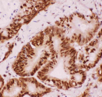
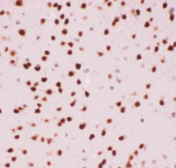
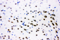
ARG42610 anti-ATF2 antibody IHC-P image
Immunohistochemistry: Paraffin-embedded Human intestinal cancer tissue. Antigen Retrieval: Heat mediation was performed in Citrate buffer (pH 6.0) for 20 min. The tissue section was blocked with 10% goat serum. The tissue section was then stained with ARG42610 anti-ATF2 antibody at 1 µg/ml dilution, overnight at 4°C.
-

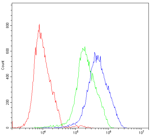
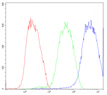
ARG42610 anti-ATF2 antibody FACS image
Flow Cytometry: HepG2 cells were blocked with 10% normal goat serum and then stained with ARG42610 anti-ATF2 antibody (blue line) at 1 µg/10^6 cells for 30 min at 20°C, followed by incubation with DyLight®488 labelled secondary antibody. Isotype control antibody (green line) was Rabbit IgG (1 µg/10^6 cells) used under the same conditions. Unlabelled sample (red line) was also used as a control.
-
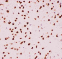
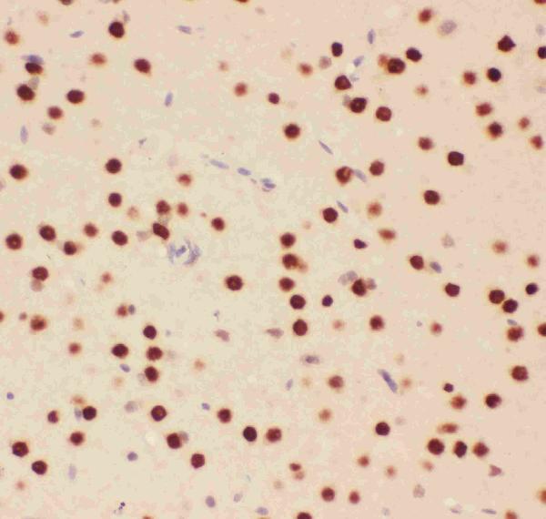
ARG42610 anti-ATF2 antibody IHC-P image
Immunohistochemistry: Paraffin-embedded Mouse brain tissue. Antigen Retrieval: Heat mediation was performed in Citrate buffer (pH 6.0) for 20 min. The tissue section was blocked with 10% goat serum. The tissue section was then stained with ARG42610 anti-ATF2 antibody at 1 µg/ml dilution, overnight at 4°C.
-
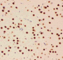
ARG42610 anti-ATF2 antibody IHC-P image
Immunohistochemistry: Paraffin-embedded Rat brain tissue. Antigen Retrieval: Heat mediation was performed in Citrate buffer (pH 6.0) for 20 min. The tissue section was blocked with 10% goat serum. The tissue section was then stained with ARG42610 anti-ATF2 antibody at 1 µg/ml dilution, overnight at 4°C.
-
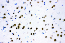
ARG42610 anti-ATF2 antibody IHC-Fr image
Immunohistochemistry: Frozen section of Rat brain tissue. The tissue section was blocked with 10% goat serum. The tissue section was then stained with ARG42610 anti-ATF2 antibody at 1 µg/ml dilution, overnight at 4°C.
-
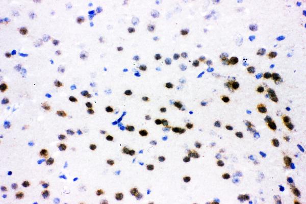
ARG42610 anti-ATF2 antibody IHC-Fr image
Immunohistochemistry: Frozen section of Mouse brain tissue. The tissue section was blocked with 10% goat serum. The tissue section was then stained with ARG42610 anti-ATF2 antibody at 1 µg/ml dilution, overnight at 4°C.
-
ARG42610 anti-ATF2 antibody FACS image
Flow Cytometry: K562 cells were blocked with 10% normal goat serum and then stained with ARG42610 anti-ATF2 antibody (blue line) at 1 µg/10^6 cells for 30 min at 20°C, followed by incubation with DyLight®488 labelled secondary antibody. Isotype control antibody (green line) was Rabbit IgG (1 µg/10^6 cells) used under the same conditions. Unlabelled sample (red line) was also used as a control.