ARG63501
anti-ASC / TMS1 antibody
anti-ASC / TMS1 antibody for IHC-Formalin-fixed paraffin-embedded sections,Western blot and Human
NLRP3 Inflammasome Study antibody; NLRC4 Inflammasome Study antibody
Overview
| Product Description | Goat Polyclonal antibody recognizes ASC / TMS1 |
|---|---|
| Tested Reactivity | Hu |
| Tested Application | IHC-P, WB |
| Specificity | This antibody is expected to recognise all reported Human isoforms according to NP_037390.2 and NP_660183.1. |
| Host | Goat |
| Clonality | Polyclonal |
| Isotype | IgG |
| Target Name | ASC / TMS1 |
| Antigen Species | Human |
| Immunogen | C-RESQSYLVEDLERS |
| Conjugation | Un-conjugated |
| Alternate Names | Apoptosis-associated speck-like protein containing a CARD; TMS; PYD and CARD domain-containing protein; hASC; ASC; TMS-1; TMS1; Caspase recruitment domain-containing protein 5; Target of methylation-induced silencing 1; CARD5 |
Application Instructions
| Application Suggestion |
|
||||||
|---|---|---|---|---|---|---|---|
| Application Note | IHC-P: Antigen Retrieval: Steam tissue section in Citrate buffer (pH 6.0). WB: Recommend incubate at RT for 1h. * The dilutions indicate recommended starting dilutions and the optimal dilutions or concentrations should be determined by the scientist. |
Properties
| Form | Liquid |
|---|---|
| Purification | Purified from goat serum by antigen affinity chromatography. |
| Buffer | Tris saline (pH 7.3), 0.02% Sodium azide and 0.5% BSA. |
| Preservative | 0.02% Sodium azide |
| Stabilizer | 0.5% BSA |
| Concentration | 0.5 mg/ml |
| Storage Instruction | For continuous use, store undiluted antibody at 2-8°C for up to a week. For long-term storage, aliquot and store at -20°C or below. Storage in frost free freezers is not recommended. Avoid repeated freeze/thaw cycles. Suggest spin the vial prior to opening. The antibody solution should be gently mixed before use. |
| Note | For laboratory research only, not for drug, diagnostic or other use. |
Bioinformation
| Database Links |
Swiss-port # Q9ULZ3 Human Apoptosis-associated speck-like protein containing a CARD |
|---|---|
| Background | ASC / TMS1 is an adaptor protein. It is composed of two protein-protein interaction domains: a N-terminal PYRIN-PAAD-DAPIN domain (PYD) and a C-terminal caspase-recruitment domain (CARD). The PYD and CARD domains are members of the six-helix bundle death domain-fold superfamily that mediates assembly of large signaling complexes in the inflammatory and apoptotic signaling pathways via the activation of caspase. In normal cells, this protein is localized to the cytoplasm; however, in cells undergoing apoptosis, it forms ball-like aggregates near the nuclear periphery. Two transcript variants encoding different isoforms have been found for this gene. [provided by RefSeq, Jul 2008] |
| Function | ASC / TMS1 functions as key mediator in apoptosis and inflammation. Promotes caspase-mediated apoptosis involving predominantly caspase-8 and also caspase-9 in a probable cell type-specific manner. Involved in activation of the mitochondrial apoptotic pathway, promotes caspase-8-dependent proteolytic maturation of BID independently of FADD in certain cell types and also mediates mitochondrial translocation of BAX and activates BAX-dependent apoptosis coupled to activation of caspase-9, -2 and -3. Involved in macrophage pyroptosis, a caspase-1-dependent inflammatory form of cell death and is the major constituent of the ASC pyroptosome which forms upon potassium depletion and rapidly recruits and activates caspase-1. In innate immune response believed to act as an integral adapter in the assembly of the inflammasome which activates caspase-1 leading to processing and secretion of proinflammatory cytokines. The function as activating adapter in different types of inflammasomes is mediated by the pyrin and CARD domains and their homotypic interactions. Required for recruitment of caspase-1 to inflammasomes containing certain pattern recognition receptors, such as NLRP2, NLRP3, AIM2 and probably IFI16. In the NLRP1 and NLRC4 inflammasomes seems not be required but facilitates the processing of procaspase-1. In cooperation with NOD2 involved in an inflammasome activated by bacterial muramyl dipeptide leading to caspase-1 activation. May be involved in DDX58-triggered proinflammatory responses and inflammasome activation. Isoform 2 may have a regulating effect on the function as inflammasome adapter. Isoform 3 seems to inhibit inflammasome-mediated maturation of interleukin-1 beta. In collaboration with AIM2 which detects cytosolic double-stranded DNA may also be involved in a caspase-1-independent cell death that involves caspase-8. In adaptive immunity may be involved in maturation of dendritic cells to stimulate T-cell immunity and in cytoskeletal rearrangements coupled to chemotaxis and antigen uptake may be involved in post-transcriptional regulation of the guanine nucleotide exchange factor DOCK2; the latter function is proposed to involve the nuclear form. Also involved in transcriptional activation of cytokines and chemokines independent of the inflammasome; this function may involve AP-1, NF-kappa-B, MAPK and caspase-8 signaling pathways. For regulation of NF-kappa-B activating and inhibiting functions have been reported. Modulates NF-kappa-B induction at the level of the IKK complex by inhibiting kinase activity of CHUK and IKBK. Proposed to compete with RIPK2 for association with CASP1 thereby down-regulating CASP1-mediated RIPK2-dependent NF-kappa-B activation and activating interleukin-1 beta processing. Modulates host resistance to DNA virus infection, probably by inducing the cleavage of and inactivating CGAS in presence of cytoplasmic double-stranded DNA (PubMed:28314590). [UniProt] |
| Highlight | Related products: TMS1 antibodies; TMS1 Duos / Panels; Anti-Goat IgG secondary antibodies; Related news: Exploring Antiviral Immune Response RIP1 activation and pathogenesis of NASH |
| Research Area | NLRP3 Inflammasome Study antibody; NLRC4 Inflammasome Study antibody |
| Calculated MW | 22 kDa |
| PTM | Phosphorylated. |
Images (3) Click the Picture to Zoom In
-
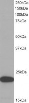
ARG63501 anti-ASC / TMS1 antibody WB image
Western blot: 35 µg of HeLa cell lysate (in RIPA buffer) stained with ARG63501 anti-ASC / TMS1 antibody at 1 µg/ml dilution and incubated at RT for 1 hour.
-
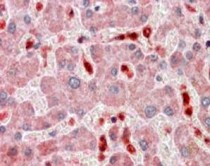
ARG63501 anti-ASC / TMS1 antibody IHC-P image
Immunohistochemistry: Paraffin-embedded Human liver tissue. Antigen Retrieval: Steam tissue section in Citrate buffer (pH 6.0). The tissue section was stained with ARG63501 anti-ASC / TMS1 antibody at 2.5 µg/ml dilution followed by AP-staining.
-
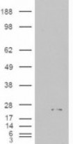
ARG63501 anti-ASC / TMS1 antibody WB image
Western blot: 1). Mock transfection; 2) ASC (RC215592) expressing plasmid transfected HEK293 cell lysate standed with ARG63501 anti-ASC / TMS1 antibody.


