ARG42964
anti-ARP2 / 3 subunit 1B antibody
anti-ARP2 / 3 subunit 1B antibody for Flow cytometry,IHC-Formalin-fixed paraffin-embedded sections,Western blot and Human,Mouse,Rat
Overview
| Product Description | Rabbit Polyclonal antibody recognizes ARP2 / 3 subunit 1B |
|---|---|
| Tested Reactivity | Hu, Ms, Rat |
| Tested Application | FACS, IHC-P, WB |
| Host | Rabbit |
| Clonality | Polyclonal |
| Isotype | IgG |
| Target Name | ARP2 / 3 subunit 1B |
| Antigen Species | Human |
| Immunogen | Recombinant protein corresponding to Q293-K372 of Human ARP2/3 subunit 1B. |
| Conjugation | Un-conjugated |
| Alternate Names | ARC41; p40-ARC; Actin-related protein 2/3 complex subunit 1B; Arp2/3 complex 41 kDa subunit; p41-ARC |
Application Instructions
| Application Suggestion |
|
||||||||
|---|---|---|---|---|---|---|---|---|---|
| Application Note | IHC-P: Antigen Retrieval: Heat mediation was performed in Citrate buffer (pH 6.0) for 20 min. * The dilutions indicate recommended starting dilutions and the optimal dilutions or concentrations should be determined by the scientist. |
||||||||
| Observed Size | ~ 40 kDa |
Properties
| Form | Liquid |
|---|---|
| Purification | Affinity purification with immunogen. |
| Buffer | 0.2% Na2HPO4, 0.9% NaCl, 0.05% Sodium azide and 4% Trehalose. |
| Preservative | 0.05% Sodium azide |
| Stabilizer | 4% Trehalose |
| Concentration | 0.5 mg/ml |
| Storage Instruction | For continuous use, store undiluted antibody at 2-8°C for up to a week. For long-term storage, aliquot and store at -20°C or below. Storage in frost free freezers is not recommended. Avoid repeated freeze/thaw cycles. Suggest spin the vial prior to opening. The antibody solution should be gently mixed before use. |
| Note | For laboratory research only, not for drug, diagnostic or other use. |
Bioinformation
| Database Links | |
|---|---|
| Gene Symbol | ARPC1B |
| Gene Full Name | actin related protein 2/3 complex, subunit 1B, 41kDa |
| Background | This gene encodes one of seven subunits of the human Arp2/3 protein complex. This subunit is a member of the SOP2 family of proteins and is most similar to the protein encoded by gene ARPC1A. The similarity between these two proteins suggests that they both may function as p41 subunit of the human Arp2/3 complex that has been implicated in the control of actin polymerization in cells. It is possible that the p41 subunit is involved in assembling and maintaining the structure of the Arp2/3 complex. Multiple versions of the p41 subunit may adapt the functions of the complex to different cell types or developmental stages. This protein also has a role in centrosomal homeostasis by being an activator and substrate of the Aurora A kinase. [provided by RefSeq, Mar 2011] |
| Function | Component of the Arp2/3 complex, a multiprotein complex that mediates actin polymerization upon stimulation by nucleation-promoting factor (NPF) (PubMed:11741539, PubMed:9230079). The Arp2/3 complex mediates the formation of branched actin networks in the cytoplasm, providing the force for cell motility (PubMed:11741539, PubMed:9230079). In addition to its role in the cytoplasmic cytoskeleton, the Arp2/3 complex also promotes actin polymerization in the nucleus, thereby regulating gene transcription and repair of damaged DNA (PubMed:29925947). The Arp2/3 complex promotes homologous recombination (HR) repair in response to DNA damage by promoting nuclear actin polymerization, leading to drive motility of double-strand breaks (DSBs) (PubMed:29925947). [UniProt] |
| Cellular Localization | Cytoplasm, cytoskeleton. [UniProt] |
| Calculated MW | 41 kDa |
Images (6) Click the Picture to Zoom In
-
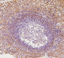
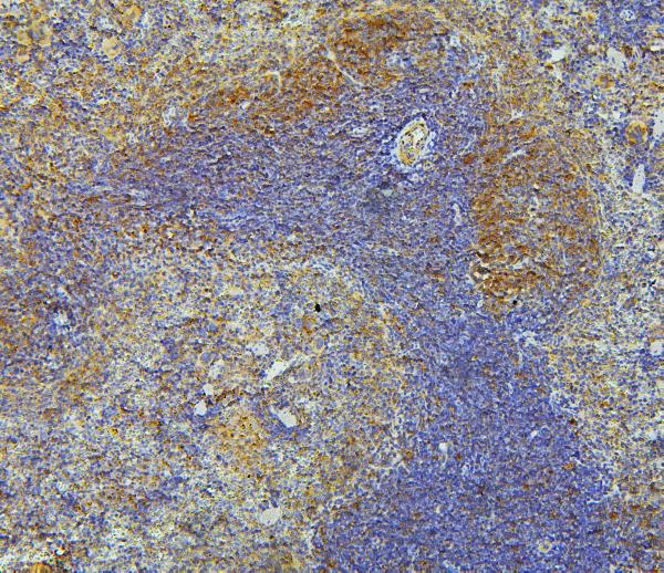
ARG42964 anti-ARP2/3 subunit 1B antibody IHC-P image
Immunohistochemistry: Paraffin-embedded Human tonsil tissue. Antigen Retrieval: Heat mediation was performed in Citrate buffer (pH 6.0) for 20 min. The tissue section was blocked with 10% goat serum. The tissue section was then stained with ARG42964 anti-ARP2/3 subunit 1B antibody at 1 µg/ml dilution, overnight at 4°C.
-

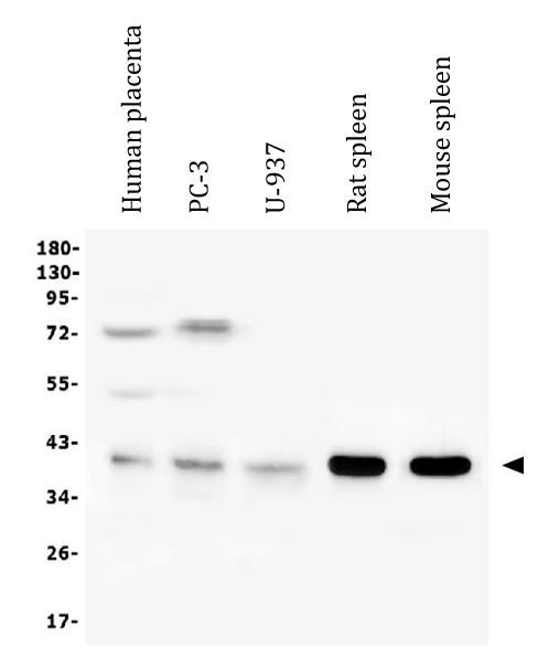
ARG42964 anti-ARP2/3 subunit 1B antibody WB image
Western blot: 50 µg of sample under reducing conditions. Human placenta, PC-3, U-937, Rat spleen and Mouse spleen lysates stained with ARG42964 anti-ARP2/3 subunit 1B antibody at 0.5 µg/ml dilution, overnight at 4°C.
-
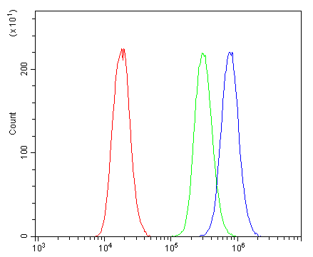
ARG42964 anti-ARP2/3 subunit 1B antibody FACS image
Flow Cytometry: PC-3 cells were blocked with 10% normal goat serum and then stained with ARG42964 anti-ARP2/3 subunit 1B antibody (blue) at 1 µg/10^6 cells for 30 min at 20°C, followed by incubation with DyLight®488 labelled secondary antibody. Isotype control antibody (green) was Rabbit IgG (1 µg/10^6 cells) used under the same conditions. Unlabelled sample (red) was also used as a control.
-
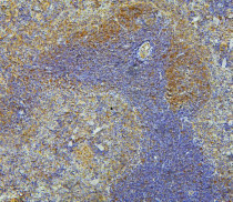
ARG42964 anti-ARP2/3 subunit 1B antibody IHC-P image
Immunohistochemistry: Paraffin-embedded Mouse spleen tissue. Antigen Retrieval: Heat mediation was performed in Citrate buffer (pH 6.0) for 20 min. The tissue section was blocked with 10% goat serum. The tissue section was then stained with ARG42964 anti-ARP2/3 subunit 1B antibody at 1 µg/ml dilution, overnight at 4°C.
-
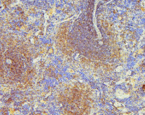
ARG42964 anti-ARP2/3 subunit 1B antibody IHC-P image
Immunohistochemistry: Paraffin-embedded Rat spleen tissue. Antigen Retrieval: Heat mediation was performed in Citrate buffer (pH 6.0) for 20 min. The tissue section was blocked with 10% goat serum. The tissue section was then stained with ARG42964 anti-ARP2/3 subunit 1B antibody at 1 µg/ml dilution, overnight at 4°C.
-

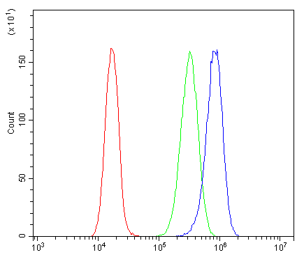
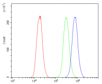
ARG42964 anti-ARP2/3 subunit 1B antibody FACS image
Flow Cytometry: CACO-2 cells were blocked with 10% normal goat serum and then stained with ARG42964 anti-ARP2/3 subunit 1B antibody (blue) at 1 µg/10^6 cells for 30 min at 20°C, followed by incubation with DyLight®488 labelled secondary antibody. Isotype control antibody (green) was Rabbit IgG (1 µg/10^6 cells) used under the same conditions. Unlabelled sample (red) was also used as a control.