ARG59541
anti-APOBEC3A / PHO1 antibody
anti-APOBEC3A / PHO1 antibody for Flow cytometry,Western blot and Human,Mouse
Overview
| Product Description | Rabbit Polyclonal antibody recognizes APOBEC3A / PHO1 |
|---|---|
| Tested Reactivity | Hu, Ms |
| Tested Application | FACS, WB |
| Host | Rabbit |
| Clonality | Polyclonal |
| Isotype | IgG |
| Target Name | APOBEC3A / PHO1 |
| Antigen Species | Human |
| Immunogen | Recombinant protein corresponding to M1-L63 of Human APOBEC3A / PHO1. |
| Conjugation | Un-conjugated |
| Alternate Names | DNA dC->dU-editing enzyme APOBEC-3A; A3A; Phorbolin-1; EC 3.5.4.- |
Application Instructions
| Application Suggestion |
|
||||||
|---|---|---|---|---|---|---|---|
| Application Note | * The dilutions indicate recommended starting dilutions and the optimal dilutions or concentrations should be determined by the scientist. |
Properties
| Form | Liquid |
|---|---|
| Purification | Affinity purification with immunogen. |
| Buffer | 0.9% NaCl, 0.2% Na2HPO4, 0.05% Sodium azide and 4% Trehalose. |
| Preservative | 0.05% Sodium azide |
| Stabilizer | 4% Trehalose |
| Concentration | 0.5 mg/ml |
| Storage Instruction | For continuous use, store undiluted antibody at 2-8°C for up to a week. For long-term storage, aliquot and store at -20°C or below. Storage in frost free freezers is not recommended. Avoid repeated freeze/thaw cycles. Suggest spin the vial prior to opening. The antibody solution should be gently mixed before use. |
| Note | For laboratory research only, not for drug, diagnostic or other use. |
Bioinformation
| Gene Symbol | APOBEC3A_B |
|---|---|
| Gene Full Name | APOBEC3A and APOBEC3B deletion hybrid |
| Background | This gene is a member of the cytidine deaminase gene family. It is one of seven related genes or pseudogenes found in a cluster, thought to result from gene duplication, on chromosome 22. Members of the cluster encode proteins that are structurally and functionally related to the C to U RNA-editing cytidine deaminase APOBEC1. The protein encoded by this gene lacks the zinc binding activity of other family members. The protein plays a role in immunity, by restricting transmission of foreign DNA such as viruses. One mechanism of foreign DNA restriction is deamination of foreign double-stranded DNA cytidines to uridines, which leads to DNA degradation. However, other mechanisms are also thought to be involved, as anti-viral effect is not dependent on deaminase activity. The protein encoded by this gene is the same as that encoded by APOBEC3A; however, this gene is a hybrid gene that results from the deletion of approximately 29.5 kb of sequence between the APOBEC3A gene and the adjacent gene APOBEC3B. The breakpoints of the deletion are within the two genes, so the deletion hybrid is predicted to have the promoter and coding region of APOBEC3A, but the 3' UTR of APOBEC3B. [provided by RefSeq, Jul 2012] |
| Function | DNA deaminase (cytidine deaminase) with restriction activity against viruses, foreign DNA and mobility of retrotransposons. Exhibits antiviral activity against adeno-associated virus (AAV) and human T-cell leukemia virus type 1 (HTLV-1) and may inhibit the mobility of LTR and non-LTR retrotransposons. Selectively targets single-stranded DNA and can deaminate both methylcytosine and cytosine in foreign DNA. Can induce somatic hypermutation in the nuclear and mitochondrial DNA. May also play a role in the epigenetic regulation of gene expression through the process of active DNA demethylation. [UniProt] |
| Cellular Localization | Nucleus. Cytoplasm. [UniProt] |
| Calculated MW | 23 kDa |
Images (2) Click the Picture to Zoom In
-
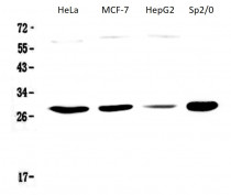
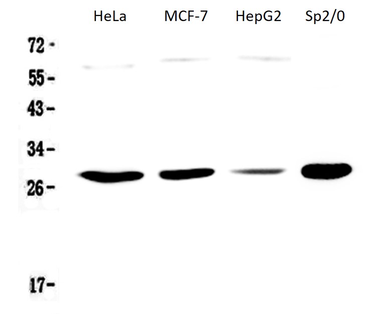
ARG59541 anti-APOBEC3A / PHO1 antibody WB image
Western blot: 50 µg of samples under reducing conditions. HeLa, MCF-7, HepG2 and Sp2/0 whole cell lysates stained with ARG59541 anti-APOBEC3A / PHO1 antibody at 0.5 µg/ml, overnight at 4°C.
-
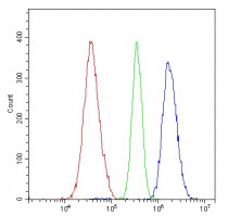
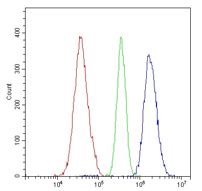
ARG59541 anti-APOBEC3A / PHO1 antibody FACS image
Flow Cytometry: A549 cells were blocked with 10% normal goat serum and then stained with ARG59541 anti-APOBEC3A / PHO1 antibody (blue) at 1 µg/10^6 cells for 30 min at 20°C, followed by incubation with DyLight®488 labelled secondary antibody. Isotype control antibody (green) was rabbit IgG (1 µg/10^6 cells) used under the same conditions. Unlabelled sample (red) was also used as a control.