ARG66617
anti-AIM2 antibody
anti-AIM2 antibody for ICC/IF,IHC-Formalin-fixed paraffin-embedded sections,Western blot and Human
Overview
| Product Description | Rabbit Polyclonal antibody recognizes AIM2 |
|---|---|
| Tested Reactivity | Hu |
| Tested Application | ICC/IF, IHC-P, WB |
| Host | Rabbit |
| Clonality | Polyclonal |
| Isotype | IgG |
| Target Name | AIM2 |
| Antigen Species | Human |
| Immunogen | KLH-conjugated synthetic peptide within the center region of Human AIM2. |
| Conjugation | Un-conjugated |
| Alternate Names | PYHIN4; Interferon-inducible protein AIM2; Absent in melanoma 2 |
Application Instructions
| Application Suggestion |
|
||||||||
|---|---|---|---|---|---|---|---|---|---|
| Application Note | IHC-P: Antigen Retrieval: Heat mediation was performed in Sodium citrate buffer (pH 6.0). * The dilutions indicate recommended starting dilutions and the optimal dilutions or concentrations should be determined by the scientist. |
||||||||
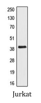
| Observed Size | ~ 39 kDa |
Properties
| Form | Liquid |
|---|---|
| Purification | Affinity purification with immunogen. |
| Buffer | 0.42% Potassium phosphate (pH 7.3), 0.87% NaCl, 0.01% Sodium azide and 30% Glycerol. |
| Preservative | 0.01% Sodium azide |
| Stabilizer | 30% Glycerol |
| Storage Instruction | For continuous use, store undiluted antibody at 2-8°C for up to a week. For long-term storage, aliquot and store at -20°C. Storage in frost free freezers is not recommended. Avoid repeated freeze/thaw cycles. Suggest spin the vial prior to opening. The antibody solution should be gently mixed before use. |
| Note | For laboratory research only, not for drug, diagnostic or other use. |
Bioinformation
| Database Links | |
|---|---|
| Gene Symbol | AIM2 |
| Gene Full Name | absent in melanoma 2 |
| Background | AIM2 is a member of the IFI20X /IFI16 family. It plays a putative role in tumorigenic reversion and may control cell proliferation. Interferon-gamma induces expression of AIM2. [provided by RefSeq, Jul 2008] |
| Function | Involved in innate immune response by recognizing cytosolic double-stranded DNA and inducing caspase-1-activating inflammasome formation in macrophages. Upon binding to DNA is thought to undergo oligomerization and to associate with PYCARD initiating the recruitment of caspase-1 precusrsor and processing of interleukin-1 beta and interleukin-18. Detects cytosolic dsDNA of viral and bacterial origin in a non-sequence-specific manner. Can also trigger PYCARD-dependent, caspase-1-independent cell death that involves caspase-8 (By similarity). Tumor suppressor which may act by repressing NF-kappa-B transcriptional activity. [UniProt] |
| Cellular Localization | Nucleus. Cytoplasm. Note=Activated inflammasomes can aggregate in the cytosol as speck-like particles. [UniProt] |
| Calculated MW | 39 kDa |
Images (3) Click the Picture to Zoom In
-
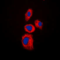
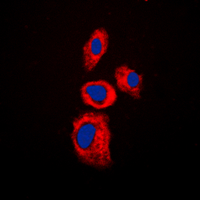
ARG66617 anti-AIM2 antibody ICC/IF image
Immunofluorescence: Formalin-fixed Jurkat cells were permeabilized with 0.1% Triton X-100 in TBS for 5-10 minutes and blocked with 3% BSA-PBS for 30 minutes at room temperature. Cells were stained with ARG66617 anti-AIM2 antibody (red) in 3% BSA-PBS and incubated overnight at 4°C. DAPI (blue) for nuclear staining.
-

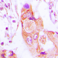
ARG66617 anti-AIM2 antibody IHC-P image
Immunohistochemistry: Formalin-fixed and paraffin-embedded Human lung cancer tissue section. Antigen Retrieval: Heat mediation was performed in Sodium citrate buffer (pH 6.0). The section was then stained with ARG66617 anti-AIM2 antibody at room temperature and detected using an HRP conjugated compact polymer system. DAB was used as the chromogen. The section was then counterstained with haematoxylin and mounted with DPX.
-
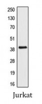
ARG66617 anti-AIM2 antibody WB image
Western blot: Jurkat whole cell lysate stained with ARG66617 anti-AIM2 antibody.