ARG66956
anti-AIF1 / Iba 1 antibody
anti-AIF1 / Iba 1 antibody for IHC-Frozen sections,IHC-Formalin-fixed paraffin-embedded sections,Western blot and Human,Mouse,Rat
Cell Biology and Cellular Response antibody; Immune System antibody; Metabolism antibody; Neuroscience antibody; Activated Macrophage/Microglia Study antibody; Neuroinflammation Study antibody; Macroglial Marker antibody
Overview
| Product Description | Rabbit Polyclonal antibody recognizes AIF1 / Iba 1 |
|---|---|
| Tested Reactivity | Hu, Ms, Rat |
| Tested Application | IHC-Fr, IHC-P, WB |
| Host | Rabbit |
| Clonality | Polyclonal |
| Isotype | IgG |
| Target Name | AIF1 / Iba 1 |
| Antigen Species | Human |
| Immunogen | Synthetic peptide around the C-terminal region of Human AIF1 / Iba1. |
| Conjugation | Un-conjugated |
| Alternate Names | Ionized calcium-binding adapter molecule 1; Allograft inflammatory factor 1; IBA1; IRT-1; IRT1; AIF-1; Protein G1 |
Application Instructions
| Application Suggestion |
|
||||||||
|---|---|---|---|---|---|---|---|---|---|
| Application Note | IHC-P: Antigen Retrieval: Boil tissue section in Sodium Citrate buffer (pH 6.0) followed by cooling at RT for 20 min. * The dilutions indicate recommended starting dilutions and the optimal dilutions or concentrations should be determined by the scientist. |
||||||||
| Positive Control | Brain tissue | ||||||||
| Observed Size | ~15 kDa |
Properties
| Form | Liquid |
|---|---|
| Purification | Affinity purified. |
| Buffer | 100 mM Tris Glycine (pH 7.0), 0.025% ProClin 300, 20% Glycerol and 1% BSA. |
| Preservative | 0.025% ProClin 300 |
| Stabilizer | 20% Glycerol and 1% BSA |
| Storage Instruction | For continuous use, store undiluted antibody at 2-8°C for up to a week. For long-term storage, aliquot and store at -20°C or below. Storage in frost free freezers is not recommended. Avoid repeated freeze/thaw cycles. Suggest spin the vial prior to opening. The antibody solution should be gently mixed before use. |
| Note | For laboratory research only, not for drug, diagnostic or other use. |
Bioinformation
| Database Links | |
|---|---|
| Gene Symbol | AIF1 |
| Gene Full Name | allograft inflammatory factor 1 |
| Background | AIF1 / Iba 1 is a protein that binds actin and calcium. This gene is induced by cytokines and interferon and may promote macrophage activation and growth of vascular smooth muscle cells and T-lymphocytes. Polymorphisms in this gene may be associated with systemic sclerosis. Alternative splicing results in multiple transcript variants, but the full-length and coding nature of some of these variants is not certain. [provided by RefSeq, Jan 2016] |
| Function | AIF1 / Iba 1 is an Actin-binding protein. It enhances membrane ruffling and RAC activation. Enhances the actin-bundling activity of LCP1. Binds calcium. Plays a role in RAC signaling and in phagocytosis. May play a role in macrophage activation and function. Promotes the proliferation of vascular smooth muscle cells and of T-lymphocytes. Enhances lymphocyte migration. Plays a role in vascular inflammation. [UniProt] |
| Highlight | Related Antibody Duos and Panels: ARG30324 Neuroinflammation Antibody Panel Related products: AIF1 antibodies; AIF1 Duos / Panels; Anti-Rabbit IgG secondary antibodies; |
| Research Area | Cell Biology and Cellular Response antibody; Immune System antibody; Metabolism antibody; Neuroscience antibody; Activated Macrophage/Microglia Study antibody; Neuroinflammation Study antibody; Macroglial Marker antibody |
| Calculated MW | 17 kDa |
| PTM | Phosphorylated on serine residues. [UniProt] |
Images (5) Click the Picture to Zoom In
-
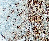
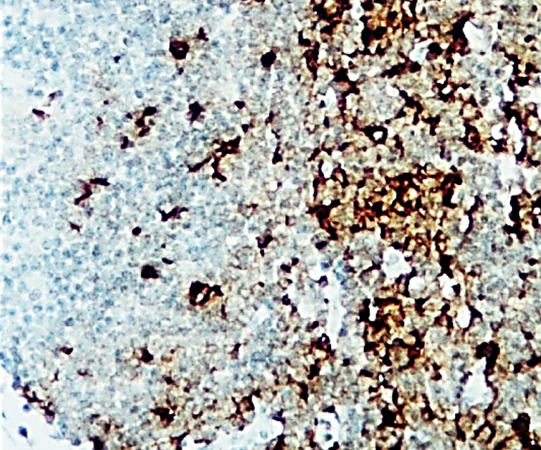
ARG66956 anti-AIF1 / Iba 1 antibody IHC-P image
Immunohistochemistry: Paraffin-embedded human lymoph node carcinoma tissue stained with ARG66956 anti-AIF1 / Iba 1 antibody at 1:200 dilution.
-
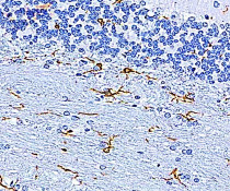
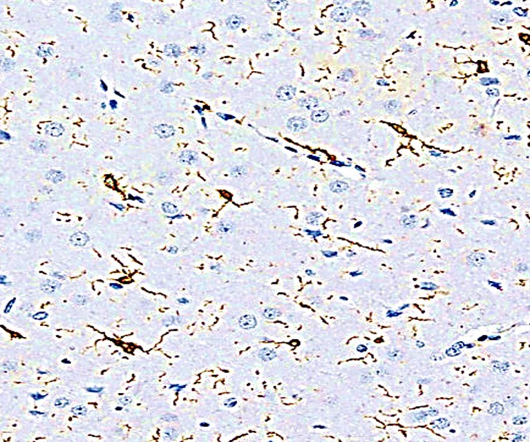
ARG66956 anti-AIF1 / Iba 1 antibody IHC-P image
Immunohistochemistry: Paraffin-embedded mouse brain tissue stained with ARG66956 anti-AIF1 / Iba 1 antibody at 1:200 dilution. Antigen Retrieval: Boil tissue section in Citrate buffer (pH 6.0).
-
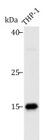
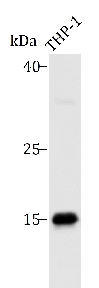
ARG66956 anti-AIF1 / Iba 1 antibody WB image
Western blot: 45 µg of THP-1 cell lysate stained with ARG66956 anti-AIF1 / Iba 1 antibody at 1:500 dilution, overnight at 4°C.
-
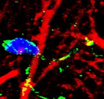
ARG66956 anti-AIF1 / Iba 1 antibody IHC-Fr image
Immunohistochemistry: Frozen section of Mouse hippocampus tissue stained with ARG66956 anti-AIF1 / Iba 1 antibody (green) at 1:200 dilution. Nuclei are stained with DAPI (blue).
-

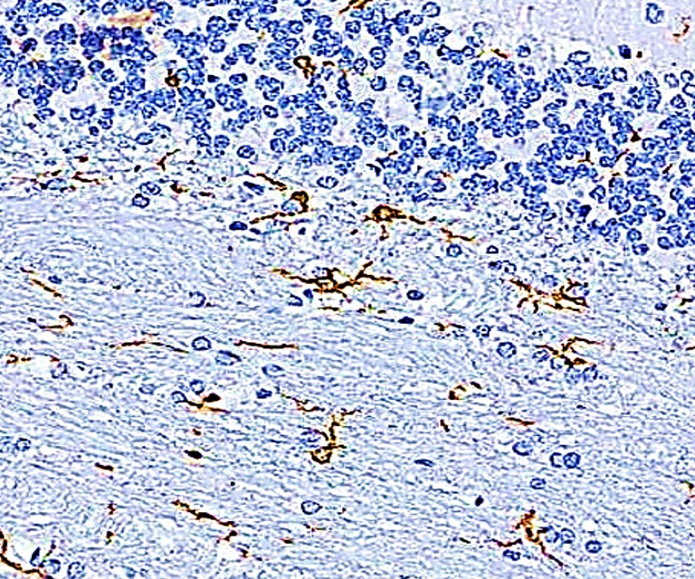
ARG66956 anti-AIF1 / Iba 1 antibody IHC-P image
Immunohistochemistry: Paraffin-embedded Rat brain tissue stained with ARG66956 anti-AIF1 / Iba 1 antibody at 1:200 dilution.