ARG63951
anti-AIF1 / Iba 1 antibody
anti-AIF1 / Iba 1 antibody for Flow cytometry,ICC/IF,IHC-Formalin-fixed paraffin-embedded sections,Western blot and Human,Mouse
Cell Biology and Cellular Response antibody; Immune System antibody; Metabolism antibody; Neuroscience antibody; Activated Macrophage/Microglia Study antibody; Neuroinflammation Study antibody; Macroglial Marker antibody
Overview
| Product Description | Goat Polyclonal antibody recognizes AIF1 / Iba 1 |
|---|---|
| Tested Reactivity | Hu, Ms |
| Predict Reactivity | Dog |
| Tested Application | FACS, ICC/IF, IHC-P, WB |
| Specificity | This antibody is expected to recognise isoform 3 (NP_001614.3) only. |
| Host | Goat |
| Clonality | Polyclonal |
| Isotype | IgG |
| Target Name | AIF1 / Iba 1 |
| Antigen Species | Human |
| Immunogen | C-NKQFLDDPKYSSDED |
| Conjugation | Un-conjugated |
| Alternate Names | Ionized calcium-binding adapter molecule 1; Allograft inflammatory factor 1; IBA1; IRT-1; IRT1; AIF-1; Protein G1 |
Application Instructions
| Application Suggestion |
|
||||||||||
|---|---|---|---|---|---|---|---|---|---|---|---|
| Application Note | IHC-P: Antigen Retrieval: Steam tissue section in Citrate buffer (pH 6.0). WB: Recommend incubate at RT for 1h. * The dilutions indicate recommended starting dilutions and the optimal dilutions or concentrations should be determined by the scientist. |
Properties
| Form | Liquid |
|---|---|
| Purification | Purified from goat serum by antigen affinity chromatography. |
| Buffer | Tris saline (pH 7.3), 0.02% Sodium azide and 0.5% BSA. |
| Preservative | 0.02% Sodium azide |
| Stabilizer | 0.5% BSA |
| Concentration | 0.5 mg/ml |
| Storage Instruction | For continuous use, store undiluted antibody at 2-8°C for up to a week. For long-term storage, aliquot and store at -20°C or below. Storage in frost free freezers is not recommended. Avoid repeated freeze/thaw cycles. Suggest spin the vial prior to opening. The antibody solution should be gently mixed before use. |
| Note | For laboratory research only, not for drug, diagnostic or other use. |
Bioinformation
| Database Links | |
|---|---|
| Background | AIF1 / Iba 1 is a protein that binds actin and calcium. This gene is induced by cytokines and interferon and may promote macrophage activation and growth of vascular smooth muscle cells and T-lymphocytes. Polymorphisms in this gene may be associated with systemic sclerosis. Alternative splicing results in multiple transcript variants, but the full-length and coding nature of some of these variants is not certain. [provided by RefSeq, Jan 2016] |
| Function | AIF1 / Iba 1 is an Actin-binding protein. It enhances membrane ruffling and RAC activation. Enhances the actin-bundling activity of LCP1. Binds calcium. Plays a role in RAC signaling and in phagocytosis. May play a role in macrophage activation and function. Promotes the proliferation of vascular smooth muscle cells and of T-lymphocytes. Enhances lymphocyte migration. Plays a role in vascular inflammation. [UniProt] |
| Research Area | Cell Biology and Cellular Response antibody; Immune System antibody; Metabolism antibody; Neuroscience antibody; Activated Macrophage/Microglia Study antibody; Neuroinflammation Study antibody; Macroglial Marker antibody |
| Calculated MW | 17 kDa |
| PTM | Phosphorylated on serine residues. |
Images (4) Click the Picture to Zoom In
-
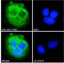
ARG63951 anti-AIF1 / Iba 1 antibody ICC/IF image
Immunofluorescence: Paraformaldehyde fixed Caco-2 cells permeabilized with 0.15% Triton. Cells were stained with ARG63951 anti-AIF1 / Iba 1 antibody (green) at 10 µg/ml dilution for 1 hour. DAPI (blue) for nuclear staining. Negative control: Unimmunized goat IgG (green) at 10 µg/ml dilution.
-
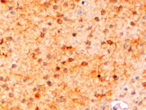
ARG63951 anti-AIF1 / Iba 1 antibody IHC-P image
Immunohistochemistry: Paraffin-embedded Mouse brain tissue. Antigen Retrieval: Steam tissue section in Citrate buffer (pH 6.0). The tissue section was stained with ARG63951 anti-AIF1 / Iba 1 antibody at 2 µg/ml dilution, followed by HRP-staining.
-
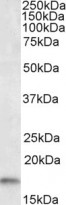
ARG63951 anti-AIF1 / Iba 1 antibody WB image
Western blot: 35 µg of Human frontal cortex lysate (in RIPA buffer) stained with ARG63951 anti-AIF1 / Iba 1 antibody at 0.5 µg/ml dilution and incubated at RT for 1 hour.
-
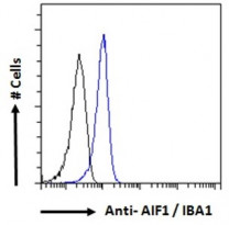
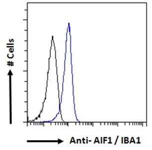
ARG63951 anti-AIF1 / Iba 1 antibody FACS image
Flow Cytometry: Paraformaldehyde-fixed K562 cells permeabilized with 0.5% Triton. Cells were stained with ARG63951 anti-AIF1 / Iba 1 antibody (blue line) at 10 µg/ml dilution for 1 hour, followed by incubation with Alexa FluorR 488 labelled secondary antibody. IgG control: Unimmunized goat IgG (black line).