ARG54374
anti-AIF antibody
anti-AIF antibody for ICC/IF,Western blot and Human
Cancer antibody; Cell Biology and Cellular Response antibody; Cell Death antibody; Gene Regulation antibody; Metabolism antibody
Overview
| Product Description | Rabbit Polyclonal antibody recognizes AIF |
|---|---|
| Tested Reactivity | Hu |
| Tested Application | ICC/IF, WB |
| Specificity | This antibody recognizes human AIF (67 kDa). |
| Host | Rabbit |
| Clonality | Polyclonal |
| Isotype | IgG |
| Target Name | AIF |
| Antigen Species | Human |
| Immunogen | Peptide corresponding to aa 109-122 at the N-terminus of human AIF. |
| Conjugation | Un-conjugated |
| Alternate Names | CMTX4; NAMSD; COWCK; Apoptosis-inducing factor 1, mitochondrial; CMT2D; EC 1.1.1.-; NADMR; PDCD8; COXPD6; AIF; Programmed cell death protein 8 |
Application Instructions
| Application Suggestion |
|
||||||
|---|---|---|---|---|---|---|---|
| Application Note | * The dilutions indicate recommended starting dilutions and the optimal dilutions or concentrations should be determined by the scientist. | ||||||
| Positive Control | K562 and Jurkat |
Properties
| Form | Liquid |
|---|---|
| Purification | Immunoaffinity chroma-tography |
| Buffer | PBS (pH 7.4) and 0.02% Sodium azide |
| Preservative | 0.02% Sodium azide |
| Storage Instruction | For continuous use, store undiluted antibody at 2-8°C for up to a week. For long-term storage, aliquot and store at -20°C or below. Storage in frost free freezers is not recommended. Avoid repeated freeze/thaw cycles. Suggest spin the vial prior to opening. The antibody solution should be gently mixed before use. |
| Note | For laboratory research only, not for drug, diagnostic or other use. |
Bioinformation
| Database Links |
Swiss-port # O95831 Human Apoptosis-inducing factor 1, mitochondrial |
|---|---|
| Gene Symbol | AIFM1 |
| Gene Full Name | apoptosis-inducing factor, mitochondrion-associated, 1 |
| Background | A novel protein that causes chromatin condensation and DNA fragmentation has been designated Apoptosis Inducing Factor (AIF). AIF localizes in mitochondria and translocates to the nucleus when apoptosis is induced. This event is followed by the release of chytochrome c and caspase-9 from mitochondria. AIF is highly conserved between human and mouse and widely expressed in different cell types and tissues. |
| Function | Functions both as NADH oxidoreductase and as regulator of apoptosis. In response to apoptotic stimuli, it is released from the mitochondrion intermembrane space into the cytosol and to the nucleus, where it functions as a proapoptotic factor in a caspase-independent pathway. In contrast, functions as an antiapoptotic factor in normal mitochondria via its NADH oxidoreductase activity. The soluble form (AIFsol) found in the nucleus induces 'parthanatos' i.e. caspase-independent fragmentation of chromosomal DNA. Interacts with EIF3G,and thereby inhibits the EIF3 machinery and protein synthesis, and activates casapse-7 to amplify apoptosis. Plays a critical role in caspase-independent, pyknotic cell death in hydrogen peroxide-exposed cells. Binds to DNA in a sequence-independent manner. [UniProt] |
| Research Area | Cancer antibody; Cell Biology and Cellular Response antibody; Cell Death antibody; Gene Regulation antibody; Metabolism antibody |
| Calculated MW | 67 kDa |
| PTM | Under normal conditions, a 54-residue N-terminal segment is first proteolytically removed during or just after translocation into the mitochondrial intermembrane space (IMS) by the mitochondrial processing peptidase (MPP) to form the inner-membrane-anchored mature form (AIFmit). During apoptosis, it is further proteolytically processed at amino-acid position 101 leading to the generation of the mature form, which is confined to the mitochondrial IMS in a soluble form (AIFsol). AIFsol is released to the cytoplasm in response to specific death signals, and translocated to the nucleus, where it induces nuclear apoptosis in a caspase-independent manner. Ubiquitination by XIAP/BIRC4 does not lead to proteasomal degradation. Ubiquitination at Lys-255 by XIAP/BIRC4 blocks its ability to bind DNA and induce chromatin degradation, thereby inhibiting its ability to induce cell death. |
Images (2) Click the Picture to Zoom In
-

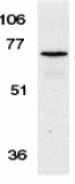
ARG54374 anti-AIF antibody WB image
Western Blot: K562 stained with ARG54374 anti-AIF antibody at 1 μg/ml dilution.
-

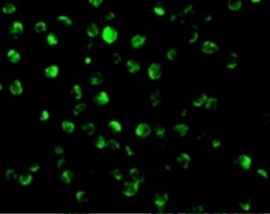
ARG54374 anti-AIF antibody ICC/IF image
Immunocytochemistry: K562 stained with ARG54374 anti-AIF antibody at 20 μg/ml dilution.