ARG41448
anti-ADRB2 antibody
anti-ADRB2 antibody for IHC-Formalin-fixed paraffin-embedded sections,Western blot and Human,Mouse,Rat
Overview
| Product Description | Rabbit Polyclonal antibody recognizes ADRB2 |
|---|---|
| Tested Reactivity | Hu, Ms, Rat |
| Tested Application | IHC-P, WB |
| Host | Rabbit |
| Clonality | Polyclonal |
| Isotype | IgG |
| Target Name | ADRB2 |
| Antigen Species | Human |
| Immunogen | Synthetic peptide of Human ADRB2. |
| Conjugation | Un-conjugated |
| Alternate Names | B2AR; BAR; BETA2AR; Beta-2 adrenergic receptor; ADRB2R; ADRBR; Beta-2 adrenoceptor; Beta-2 adrenoreceptor |
Application Instructions
| Application Suggestion |
|
||||||
|---|---|---|---|---|---|---|---|
| Application Note | IHC-P: Antigen Retrieval: Heat mediation was performed in Citrate buffer (pH 6.0, epitope retrieval solution) for 20 min. * The dilutions indicate recommended starting dilutions and the optimal dilutions or concentrations should be determined by the scientist. |
||||||
| Positive Control | A431 | ||||||
| Observed Size | ~ 68 kDa |
Properties
| Form | Liquid |
|---|---|
| Purification | Affinity purified. |
| Buffer | PBS (pH 7.4), 150 mM NaCl, 0.02% Sodium azide and 50% Glycerol. |
| Preservative | 0.02% Sodium azide |
| Stabilizer | 50% Glycerol |
| Storage Instruction | For continuous use, store undiluted antibody at 2-8°C for up to a week. For long-term storage, aliquot and store at -20°C. Storage in frost free freezers is not recommended. Avoid repeated freeze/thaw cycles. Suggest spin the vial prior to opening. The antibody solution should be gently mixed before use. |
| Note | For laboratory research only, not for drug, diagnostic or other use. |
Bioinformation
| Database Links | |
|---|---|
| Gene Symbol | ADRB2 |
| Gene Full Name | adrenoceptor beta 2, surface |
| Background | This gene encodes beta-2-adrenergic receptor which is a member of the G protein-coupled receptor superfamily. This receptor is directly associated with one of its ultimate effectors, the class C L-type calcium channel Ca(V)1.2. This receptor-channel complex also contains a G protein, an adenylyl cyclase, cAMP-dependent kinase, and the counterbalancing phosphatase, PP2A. The assembly of the signaling complex provides a mechanism that ensures specific and rapid signaling by this G protein-coupled receptor. This gene is intronless. Different polymorphic forms, point mutations, and/or downregulation of this gene are associated with nocturnal asthma, obesity and type 2 diabetes. [provided by RefSeq, Jul 2008] |
| Function | Beta-adrenergic receptors mediate the catecholamine-induced activation of adenylate cyclase through the action of G proteins. The beta-2-adrenergic receptor binds epinephrine with an approximately 30-fold greater affinity than it does norepinephrine. [UniProt] |
| Cellular Localization | Cell membrane; Multi-pass membrane protein. Early endosome. Note=Colocalizes with VHL at the cell membrane (PubMed:19584355). Activated receptors are internalized into endosomes prior to their degradation in lysosomes (PubMed:20559325). [UniProt] |
| Calculated MW | 46 kDa |
| PTM | Palmitoylated; may reduce accessibility of Ser-345 and Ser-346 by anchoring Cys-341 to the plasma membrane. Agonist stimulation promotes depalmitoylation and further allows Ser-345 and Ser-346 phosphorylation. Phosphorylated by PKA and BARK upon agonist stimulation, which mediates homologous desensitization of the receptor. PKA-mediated phosphorylation seems to facilitate phosphorylation by BARK. Phosphorylation of Tyr-141 is induced by insulin and leads to supersensitization of the receptor. Polyubiquitinated. Agonist-induced ubiquitination leads to sort internalized receptors to the lysosomes for degradation (PubMed:19424180, PubMed:20559325). Deubiquitination by USP20 and USP33, leads to ADRB2 recycling and resensitization after prolonged agonist stimulation. USP20 and USP33 are constitutively associated and are dissociated immediately after agonist stimulation. Ubiquitination by the VHL-E3 ligase complex is oxygen-dependent. Hydroxylation by EGLN3 occurs only under normoxia and increases the interaction with VHL and the subsequent ubiquitination and degradation of ADRB2. [UniProt] |
Images (2) Click the Picture to Zoom In
-
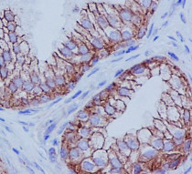
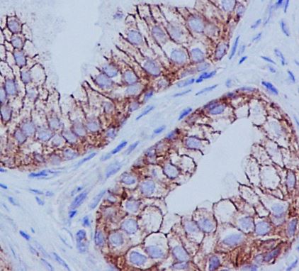
ARG41448 anti-ADRB2 antibody IHC-P image
Immunohistochemistry: Paraffin-embedded Human prostate tissue. Antigen Retrieval: Heat mediation was performed in Citrate buffer (pH 6.0, epitope retrieval solution) for 20 min. The tissue section was blocked with 10% goat serum. The tissue section was then stained with ARG41448 anti-ADRB2 antibody at 1 µg/ml dilution, overnight at 4°C.
-
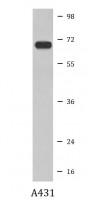
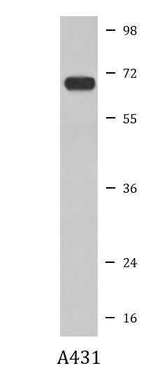
ARG41448 anti-ADRB2 antibody WB image
Western blot: A431 cell lysate stained with ARG41448 anti-ADRB2 antibody.