ARG43392
anti-ACVRL1 antibody
anti-ACVRL1 antibody for Flow cytometry,IHC-Formalin-fixed paraffin-embedded sections,Western blot and Human,Mouse,Rat
Overview
| Product Description | Rabbit Polyclonal antibody recognizes ACVRL1 |
|---|---|
| Tested Reactivity | Hu, Ms, Rat |
| Tested Application | FACS, IHC-P, WB |
| Host | Rabbit |
| Clonality | Polyclonal |
| Isotype | IgG |
| Target Name | ACVRL1 |
| Antigen Species | Human |
| Immunogen | Recombinant protein corresponding to T271-L362 of Human ACVRL1. |
| Conjugation | Un-conjugated |
| Alternate Names | ACVRLK1; ALK1; ORW2; ALK-1; HHT; EC 2.7.11.30; Serine/threonine-protein kinase receptor R3; TGF-B superfamily receptor type I; HHT2; SKR3; TSR-I; Activin receptor-like kinase 1 |
Application Instructions
| Application Suggestion |
|
||||||||
|---|---|---|---|---|---|---|---|---|---|
| Application Note | IHC-P: Antigen Retrieval: Heat mediation was performed in EDTA buffer (pH 8.0). * The dilutions indicate recommended starting dilutions and the optimal dilutions or concentrations should be determined by the scientist. |
||||||||
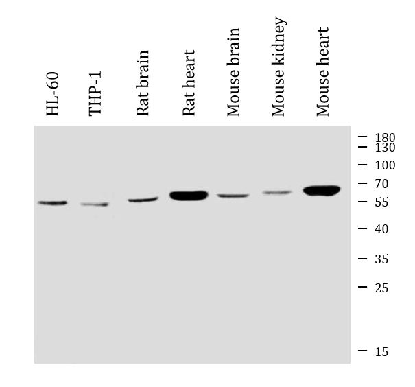
| Observed Size | 55 ~ 60 kDa |
Properties
| Form | Liquid |
|---|---|
| Purification | Affinity purification with immunogen. |
| Buffer | 0.2% Na2HPO4, 0.9% NaCl and 4% Trehalose. |
| Stabilizer | 4% Trehalose |
| Concentration | 0.5 mg/ml |
| Storage Instruction | For continuous use, store undiluted antibody at 2-8°C for up to a week. For long-term storage, aliquot and store at -20°C or below. Storage in frost free freezers is not recommended. Avoid repeated freeze/thaw cycles. Suggest spin the vial prior to opening. The antibody solution should be gently mixed before use. |
| Note | For laboratory research only, not for drug, diagnostic or other use. |
Bioinformation
| Database Links | |
|---|---|
| Gene Symbol | ACVRL1 |
| Gene Full Name | activin A receptor type II-like 1 |
| Background | This gene encodes a type I cell-surface receptor for the TGF-beta superfamily of ligands. It shares with other type I receptors a high degree of similarity in serine-threonine kinase subdomains, a glycine- and serine-rich region (called the GS domain) preceding the kinase domain, and a short C-terminal tail. The encoded protein, sometimes termed ALK1, shares similar domain structures with other closely related ALK or activin receptor-like kinase proteins that form a subfamily of receptor serine/threonine kinases. Mutations in this gene are associated with hemorrhagic telangiectasia type 2, also known as Rendu-Osler-Weber syndrome 2. [provided by RefSeq, Jul 2008] |
| Function | Type I receptor for TGF-beta family ligands BMP9/GDF2 and BMP10 and important regulator of normal blood vessel development. On ligand binding, forms a receptor complex consisting of two type II and two type I transmembrane serine/threonine kinases. Type II receptors phosphorylate and activate type I receptors which autophosphorylate, then bind and activate SMAD transcriptional regulators. May bind activin as well. [UniProt] |
| Cellular Localization | Cell membrane; Single-pass type I membrane protein. [UniProt] |
| Calculated MW | 56 kDa |
Images (6) Click the Picture to Zoom In
-
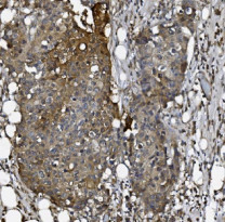
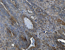
ARG43392 anti-ACVRL1 antibody IHC-P image
Immunohistochemistry: Paraffin-embedded Human breast cancer tissue. Antigen Retrieval: Heat mediation was performed in EDTA buffer (pH 8.0). The tissue section was blocked with 10% goat serum. The tissue section was then stained with ARG43392 anti-ACVRL1 antibody at 2 µg/ml dilution, overnight at 4°C.
-
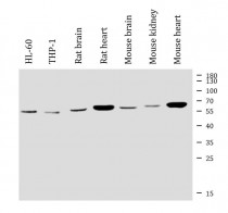
ARG43392 anti-ACVRL1 antibody WB image
Western blot: 30 µg of sample under reducing conditions. HL-60, THP-1, Rat brain, Rat heart, Mouse brain, Mouse kidney and Mouse heart lysates stained with ARG43392 anti-ACVRL1 antibody at 0.5 µg/ml dilution, overnight at 4°C.
-
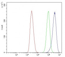
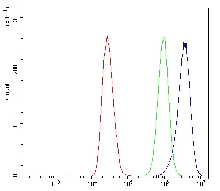
ARG43392 anti-ACVRL1 antibody FACS image
Flow Cytometry: MCF7 cells were blocked with 10% normal goat serum and then stained with ARG43392 anti-ACVRL1 antibody (blue) at 1 µg/10^6 cells for 30 min at 20°C, followed by incubation with DyLight®488 labelled secondary antibody. Isotype control antibody (green) was rabbit IgG (1 µg/10^6 cells) used under the same conditions. Unlabelled sample (red) was also used as a control.
-
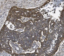
ARG43392 anti-ACVRL1 antibody IHC-P image
Immunohistochemistry: Paraffin-embedded Human gallbladder adenocarcinoma tissue. Antigen Retrieval: Heat mediation was performed in EDTA buffer (pH 8.0). The tissue section was blocked with 10% goat serum. The tissue section was then stained with ARG43392 anti-ACVRL1 antibody at 2 µg/ml dilution, overnight at 4°C.
-

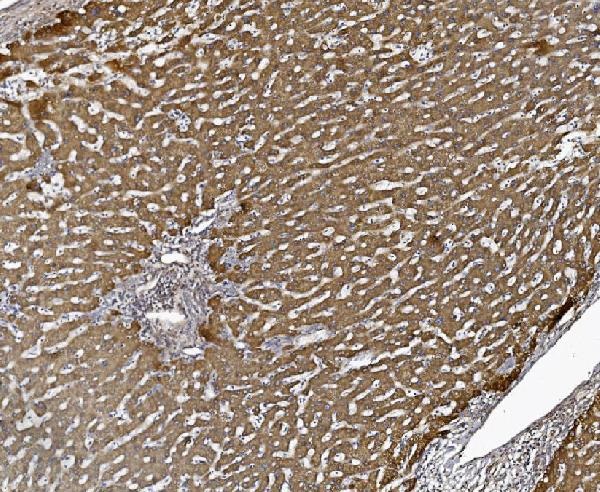
ARG43392 anti-ACVRL1 antibody IHC-P image
Immunohistochemistry: Paraffin-embedded Human liver cancer tissue. Antigen Retrieval: Heat mediation was performed in EDTA buffer (pH 8.0). The tissue section was blocked with 10% goat serum. The tissue section was then stained with ARG43392 anti-ACVRL1 antibody at 2 µg/ml dilution, overnight at 4°C.
-
ARG43392 anti-ACVRL1 antibody IHC-P image
Immunohistochemistry: Paraffin-embedded Human ovarian serous adenocarcinoma tissue. Antigen Retrieval: Heat mediation was performed in EDTA buffer (pH 8.0). The tissue section was blocked with 10% goat serum. The tissue section was then stained with ARG43392 anti-ACVRL1 antibody at 2 µg/ml dilution, overnight at 4°C.